
Journal of Clinical Images and Medical Case Reports
ISSN 2766-7820
Clinical Image - Open Access, Volume 5
A male with urinary bladder giant stone
José Richard Tenazoa Villalobos1,3*; Edgar Fermín Yan Quiroz2,3
1Hospital Víctor Lazarte Echegaray – Es Salud, Trujillo 13006, Peru.
2Virgen de la Puerta High Complexity Hospital – Es Salud, La Esperanza 13013, Peru.
3Faculty of Medicine, Universidad Privada Antenor Orrego, Trujillo 13008, Peru.
*Corresponding Author : Villalobos JRT
Hospital Víctor Lazarte Echegaray – EsSalud, Trujillo
13006, Peru.
Email: shinato_fenix@hotmail.com
Received : Feb 05, 2024
Accepted : Feb 19, 2024
Published : Feb 26, 2024
Archived : www.jcimcr.org
Copyright : © Villalobos JRT (2024).
Abstract
Citation: Villalobos JRT, Quiroz EFY. A male with urinary bladder giant stone. J Clin Images Med Case Rep. 2024; 5(2): 2881.
Description
86 years old male patient with antecedent of traumatic fracture of the spine and paraparesis state, 2 months ago presents dysuria, frecuency, vesical tenesmus, urinary retention and progression to anuria associated to abdominopelvic pain with irradiation to the lumbar zone. At emergency department there are many failed intents of urinary bladder catheterization and no relieved of pain (Figure 1). Figure 2 shows an ovoid concentric, multilayer and hyperdense mass in the pelvis. Serum creatinine: 4 mg/dl, serum ureic nitrogen: 89 mg/dl.
A few hours after, patient was evaluated by the Urologist and went to the operation room and founded a stone inside the urinary bladder who acquire its form, the complete remotion of the calculi was through an incision, remotion of the bladder was no necessary, the patient had a favorable evolution, and the urinary function was restored, the surgery was successfully and discharge one week after.
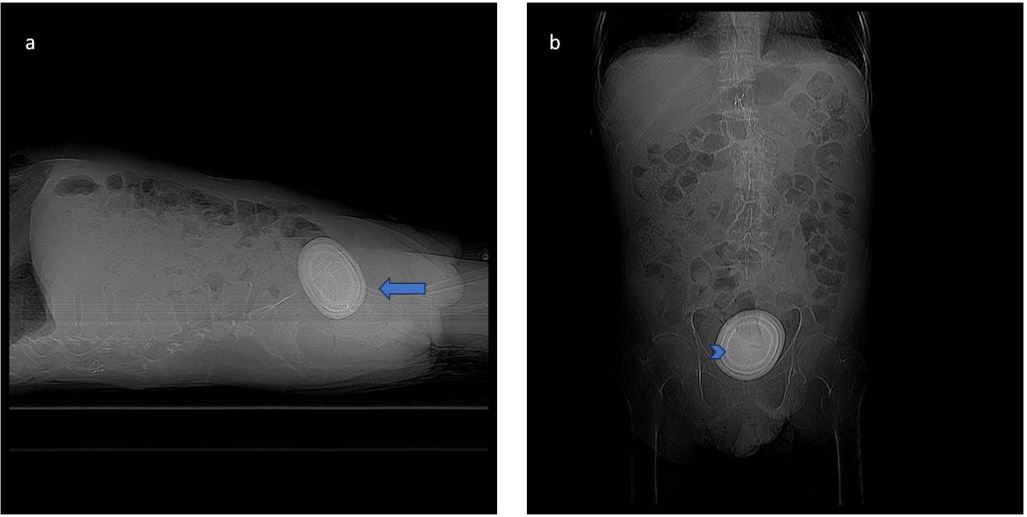
a. Lateral x-ray abdomen study reveal a ovoid mass with multiple concentric lines in layer pattern (arrow) in the pelvis, that correspond to a caculi with form of urinary bladder.
b. Anteroposterior x – ray abdomen study show the inner layer (arrowhead).
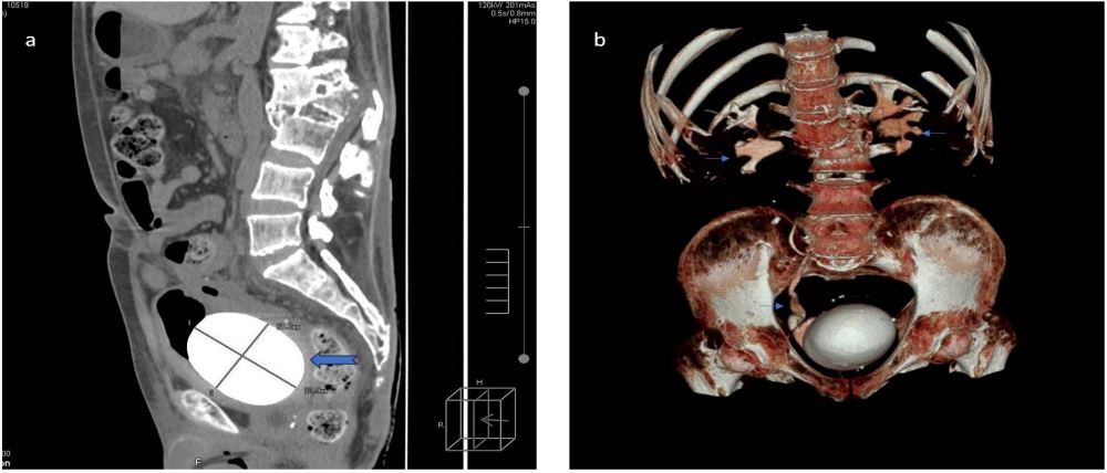
b. Urothomography and reconstruction reveal a mass inside the urinary bladder with the rigth ureter dilated and bilateral hydronephrosis (thin arrow).
Declarations
Conflicts of interest: The authors declare that they have no financial or non-financial conflicts of interest in the publication of this article.
Financing statement: This project was self-funded by the authors and did not receive funding from any external source.
Author contributions: All authors have contributed to, read and approved the final manuscript for submission José Richard Tenazoa Villalobos: Patient care and manuscript writing. Edgar Fermín Yan-Quiroz: Patient care and manuscript writing.