
Journal of Clinical Images and Medical Case Reports
ISSN 2766-7820
Case Report - Open Access, Volume 5
Effects of photobiomodulation on wound dehiscence in oncology: A case report
El Hadioui Mohamed Reda1*; Camelia Billard-Sandu2; Nahla Belattar3; Andreea Cabulca3; Layal Rached3; Leony Antoun4
1Cadi Ayyad University, Internship Rotation at Gustave Roussy, Oncology Hospital, France.
2Chief of Department, Indre et Loire -Gustave Roussy, France.
3Indre et Loire Department, France.
4Department of Clinical Oncology and Hematology -Gustave Roussy, France.
*Corresponding Author : El Hadioui M Reda
Student, Cadi Ayyad University, Internship Rotation
at Gustave Roussy, Oncology Hospital, France.
Tel: +33 629260986.
Email: mohamedredaelhadioui@gmail.com
Received : Feb 03, 2024
Accepted : Feb 21, 2024
Published : Feb 28, 2024
Archived : www.jcimcr.org
Copyright : © Reda EHM (2024).
Abstract
Introduction: Wound healing is a defined physiological process which depends on multiple factors that can be lacking in cancerous patients. Reopening, infection and/or hyperscarification can be a common consequence of pathological cicatrisation. To counter them, therapists may use antibiotics, surgery, or laser. Photobiomodulation is considered to be one of the techniques used in tissue healing, inflammation, and pain relief.
Case presentation: We report the case of a 53-year-old female patient who had a post operative deep inferior epigastric perforator DIEP flap surgery wound reopening in the context of her breast cancer surgery; photobiomodulation ATP38 was used to treat the infected wound following the protocol of 15 sessions, spaced by 2 days. The parameters used were fluence max 4J/m2 , 70W, in a continuous mode, 470 nm emission followed by 620 nm.
Conclusion: Photobiomodulation has demonstrated a remarkable improvement in our case. Low-level laser therapy may offer a good alternative for wound healing recovery in the context of cancer.
Keywords: Photobiomodulation; Scar; Infection; Cancer; Surgery.
Citation: Reda EHM, Billard-Sandru C, Belattar N, Cabulca A, Rached L, et al. Effects of photobiomodulation on wound dehiscence in oncology: A case report. J Clin Images Med Case Rep. 2024; 5(2): 2885.
Introduction
Abdominal wound dehiscence is defined as a separation of layers of the abdominal wall. Despite the advances in surgery and the post operative care, the mortality related to wound dehiscence has not improved [1]. The etiology behind it can be a consequence of local to systemic factors such as diabetes, pulmonary disease, dynamic instability, wound infection,metabolic disorders, the use of steroids and others. In the case of cancer, the immunocompromised state of health can be an additional risk factor to develop a wound reopening. The treatments can vary from local topical treatments to systemic injections. In our case the decision was to start a low-level laser therapy to simulate locally and promote better scar healing. Photobiomodulation PBM, also known as low-level laser therapy, was discovered 50 years ago by Ender master in Hungary. It has indications in the medical field as well as dentistry and cosmetics. In the recent few years there has been a major progress in studies, trying to understand the actual mechanism of it; the most generic explanation of being the stimulation of the chromophores and specifically, the fourth unit of the mitochondrial electron transport chain [2]. The concept of the treatment is based on the wavelength.The absorption of light by cytochrome c-oxidase located in the fourth unit of the respiratory chain results in the activation of the proton gradient. Consequently, an increase of ATP production levels leads to reactivation of oxygen species and calcium ions. All of the metabolic reactions or pathways lead to the differentiation, proliferation, migration of cells resulting in an increase of protein production, collagen simulation, and scarring tissue. Medically speaking wound healing after laparotomy requires oxygen consumption, the absence of toxic or septic factors and normal glycemia which can be disregulated in the field of tumorous states of health. Therefore, the aim of this paper is to present a clinical case based on a review of low level laser therapy use for the wound dehiscence particularly in cancerous patient.
Case report
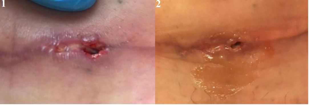
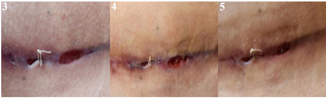
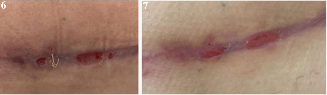
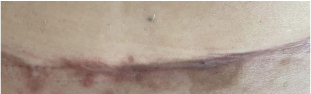
A 53-year-old patient was addressed to radiotherapy department for an infected wound reopening a week after deep inferior epigastric perforator DIEP flap surgery in the context of breast cancer. The history of the disease consists of breast cancer diagnosed at screening in March 2018 to which the patient received a neoadjuvant chemotherapy, followed by a tumorectomy. A local relapse at screening was discovered in July 2023, for which the patient received a mastectomy, lymph node dissection, and DIEP flap surgery, followed by adjuvant chemotherapy. She consulted a few days following surgery class I according to SWC [4], after noticing a wound reopening of 3 cm and 1 cm in the hypogastric region. Clinical examination showed a stable patient with signs of local infection. Her pain score was 1/10. The pelvic exam showed 3 cm and 1 cm opening with inflammatory local symptoms (redness, heat, oozing pus) (Figure 2). The patient was seen by her surgeon who prescribed antibiotics amoxicillin clavulanate at dose of 3 g/day for a duration of seven days which resulted in the wound drying without subjective improvement regarding the reopening as proved by sequential pictures taken by the patient and later on by the medical staff (Figure 3 end of antibiotics). The surgeon decided to put a stitch in end of October, post poning her first chemotherapy session. The patient started her first chemotherapy session by the end of October (Figure 3). Considering the fluctuation of the local dehiscence during the month of November (progession and regression of the healing process), the decision at radiotherapy department was to start low-level laser therapy after informing the patient of the side effects, contraindications of PBM in a context of shared decision making. The protocol that was followed, was “complicated wound” ATP38 blue light wavelength of (470 nm) for 575 seconds, followed by “curative epithelitis” ATP38 of red light wavelength 620 nm for 488 seconds at an energy density of 4j/m2 and a frequency of 70Hz. The sessions were implemented for 3 weeks spaced by 48 hours for a total treatment of 15 sessions. The change of the wound went from wound dehiscence stage (Figure 6) to wound closure (Figure 7) to healed scar (Figure 8).
Discussion
The positive evolution of the tissue healing while following a PBM protocol makes us consider its implication in helping patients with their scarring process. What’s particular about this case is that the reopening happened in the context of local infection treated by antibiotics with a delayed scar healing (2 months time). During the sessions, the patient experienced no additional side effect and was notified to pay attention to any possible unwanted outcomes of the PBM. When laser was used, not only the wounds stopped oozing which was noticed by constant clinical examination throughout the sessions, but also we witnessed a progressive closure of the reopening (less than 3 weeks, considering the wound was not ameliorating for more than 2 months as reported by the patient). These results makes us think about the contribution of laser in the context of cancer outcomes, especially with patients whose biological markers show that the inflammatory cells cannot counter infection or dermatological disorders. Therefore, the local simulation seems to be a very interesting perspective that goes hand in hand with scar healing. A recent study showed that there is a duality in the use of PBM, as if the wavelength reaches a maximum level, the effect on the cells becomes more decreasing, then increasing [6]. Thus, this should be taken into consideration within the protocol in order to be beneficial and not harmful to the patient, as well as developing more tools and guidelines to ensure the good use of PBM. The use of chemotherapy and radiotherapy is very common for cancer patients which indefinitely leads to side effects such as mucositis, dysphasia, dyslexia, dermatitis, hypo salivation, lymphoedema, and others which can be prevented by the use of PBM, nevertheless, it’s indication in wound dehiscence is not shed light on in oncology. We must acknowledge the fact that PBM requires several sessions to show great results, but it’s non-invasive and non-painful, short timed procedure makes it medically acceptable by patients.
Furthermore, regarding our case, wound dehiscence delayed the start of chemotherapy sessions which is a good argument to investigate the medical alternatives that can possibly reduce pathological woundhealing, and thus have an indirect impact on the efficacy of oncological treatments. Overall, further clinical studies are still necessary to work on the mechanisms of PBM, as well as its vast indication pool that can not only make acute changes such as in mucositis, but also in the long-term by analysing its effect on enhancing the quality of life for cancer patients Qo l as well as preventing side effects in the field of oncology. And by that we mean, chemotherapy’s side effects in the neurological aspects, radiotherapy in the dermatological/ ENT aspects, and carcinologic surgeries as its the case of our study.
Conclusion
Practitioners in the field of oncology should consider photobiomodulation in the case of wound dehiscence as it has demonstrated a remarkable improvement in our case. Low-level laser therapy may offer a good alternative for preventing and positively enhancing wound healing process in the context of cancer.
Ethical considerations: Informed consent was obtained from the patient for publication this report.
References
- Spiliotis J, Tsiveriotis K, Datsis AD. et al. Wound dehiscence: is still a problem in the 21th century: a retrospective study. World J Emerg Surg. 2009; 4: 12.
- Dompe C, Moncrieff L, Matys J, Grzech-Leśniak K, Kocherova I, Bryja A, Bruska M, Dominiak M, Mozdziak P, Skiba THI, et al. Photobiomodulation—Underlying Mechanism and Clinical Applications. Journal of Clinical Medicine. 2020; 9(6): 1724.
- Barasch A, Migliorati CA, Milstein, DM, Genot MT, Lansaat L, van der Brink R, et al. Lowlevel laser therapy/photobiomodulation in the management of side effects of chemoradiation therapy in head andneck cancer: part 1: mechanisms of action, dosimetric, and safety considerations. Supportive care in cancer: official journal of the Multinationalassociation of Supportive Care in Cancer, 2016; 24(6): 2781-2792.
- Herman TF, Bordoni B. Wound Classification. [Updated 2023 Aug 17]. In: StatPearls [Internet]. Treasure Island (FL): StatPearls Publishing. 2024.
- Mester E, Spiry T, Szende B, Tota JG. Effect of laser rays on wound healing. Am J Surg. 1971; 122: 532-535.
- Huang YY, Sharma SK, Carroll JD, et al. Biphasic dose response in low level light therapy—an update. Dose Response. 2011; 9: 602-618.
- X.Araújo JGL, Araújo EMDS, Rodrigues FCN, Paschoal MAB, Lago ADN. High power laser and photobiomodulation in oral surgery: case report. J Lasers Med Sci. 2019; 10(1): 75-78. doi:10.15171/jlms.2019.12.
- Michael R Hamblin. Mechanisms and applications of the antiinflammatory effects of photobiomodulation[J]. AIMS Biophysics. 2017; 4(3): 337-361. doi: 10.3934/biophy.2017.3.337.
- Jabłoński P, Musiał M, Wiench R, Stefanik N, Olchowy C, Matys J, Skaba D, GrzechLeśniak K. Photobiomodulation Therapy in the Treatment of Oral Mucositis—A Case Report. Medicina. 2022; 58(5): 618. https://doi.org/10.3390/medicina58050618.
- Barolet D, Roberge CJ, Auger FA, Boucher A, Germain L. Regulation of skin collagen metabolism in vitro using a pulsed 660 nm LED light source: clinical correlation with a single-blinded study. J Invest Dermatol. 2009; 129(12): 2751-9. doi: 10.1038/ jid.2009.186. Epub 2009 Jul 9. PMID: 19587693.