
Journal of Clinical Images and Medical Case Reports
ISSN 2766-7820
Case Report - Open Access, Volume 5
Symmetrical peripheral gangrene: A rare case with unpredictable presentation and tragic outcome
Rameez Hussain1*; Aleena Aftab1 ; Sakina Abbas1 ; Mansoor Ahmed1 ; Anum Naqvi1 ; Radeyah Waseem1 ; Afnan WM Jobran2 ; Hussain Haider Shah1 ; Tirth Dave3
1Department of Medicine, Dow University of Health Sciences, Pakistan.
2Department of Medicine, Al-Quds University, Jerusalem, Palestine.
3Department of Medicine, Bukovinian State Medical University, Chernivtsi, Ukraine.
*Corresponding Author : Rameez Hussain
Department of Medicine, Dow University of Health
Sciences, Pakistan.
Email: rameezh289@gmail.com
Received : Feb 21, 2024
Accepted : Mar 08, 2024
Published : Mar 15, 2024
Archived : www.jcimcr.org
Copyright : © Hussain R (2024).
Abstract
Symmetrical peripheral gangrene (SPG) is a rare condition characterized by acute ischemic damage in multiple extremities without any obstruction or vasculitis in the supplying vessels. It commonly affects the toes, hands, scrotum, and earlobes, increasing the risk of limb amputation and significantly impacting the patient’s quality of life. The underlying vascular injury mechanism is disseminated intravascular coagulation, and SPG can manifest unpredictably in conditions associated with septic shock, low output states, vasospastic conditions, myeloproliferative disorders, or hyper viscosity syndrome. In this case study, a 19-year-old woman presented with bluish-black blisters on her feet and legs, accompanied by pain. The blisters gradually increased in size and burst, resulting in a watery discharge tinged with blood. Despite various investigations and treatments involving multiple departments, the patient ultimately succumbed to septic shock and passed away on June 13, 2023, as confirmed by a house officer and a senior resident.
Keywords: Symmetrical peripheral gangrene; Disseminated intravascular coagulation; Purpura fulminans; Vascular occlusive disease; Civil Hospital Karachi.
Citation: Hussain R, Aftab A, Abbas S, Ahmed M, Naqvi A et.al. Symmetrical peripheral gangrene: A rare case with unpredictable presentation and tragic outcome. J Clin Images Med Case Rep. 2024; 5(3): 2923.
Introduction
Symmetrical peripheral gangrene (SPG) also known as purpura fulminans is a rare clinical syndrome characterized by acute onset of symmetrical ischemic damage, which occurs at the distal part of the limbs or genitalia in the absence of a major vascular occlusive disease. It imposes an elevated risk of mortality and high frequency of multiple limb amputation in up to 70% of patients who survive [1]. The most affected body parts include the toes, hands, scrotum, and earlobes. It is a rare but severe complication of disseminated intravascular coagulation (DIC) often accompanied by sepsis [2,3]. There are only a few cases of SPG reported worldwide including South Asian countries such as India and Pakistan. A thorough research in the literature revealed that only two cases of SPG have been reported in Pakistan, both of which were associated with postpartum septic shock [4,5]. Here we report a case of 19 years old female who presented to OPD of Civil Hospital, Karachi with complaints of blisters on bilateral foot and bilateral foot discoloration extending to legs for 15 days.
Case presentation
A 19-year-old woman, previously in her usual state of health, initiated her medical journey when she experienced a sudden onset of perplexing symptoms. Fifteen days before her admission, she noticed the emergence of bluish-black blisters on both of her feet. The lesions were accompanied by an unsettling discoloration that extended towards her legs. Alarmed by the persistence and progression of her symptoms, she sought medical attention and presented it to the outpatient department on March 31st. These blisters, measuring around 3-4 cm, were not only physically discomforting but also emotionally distressing as they became a source of moderate to severe pain. Their alarming trajectory unfolded as the blisters grew over time, ultimately rupturing to release a watery discharge tinged with blood. Curiously, the patient adamantly denied any history of trauma, fever, or cold-induced pain in her toes, making the origin of her condition even more baffling. Furthermore, she disclosed no signs of discomfort related to alopecia, oral ulcers, joint pain, or a rash triggered by sunlight. Though a diligent student, she did not report any notable familial medical conditions.
A thorough examination upon admission on April 1st unveiled a mix of puzzling and concerning clinical findings. Her body temperature remained within the normal range, while her blood pressure was recorded at 130/80 mmHg. The respiratory rate stood at 20 breaths per minute, and the pulse rate was measured at 110 beats per minute. Notably, her sub-vital analysis pointed towards anemia. Local examination revealed striking discoloration and substantial swelling in both lower limbs, encompassing an area that extended up to her thighs. The right-sided toenails were notably blackened, accompanied by multiple fluid-filled blisters present on both lower limbs. Remarkably, the largest blister measured approximately 8 cm, underscoring the severity of her condition. While the pulses in her femoral artery were palpable on both sides, the popliteal pulse exhibited weakness, and the dorsal pedis pulse was conspicuously absent. Additionally, the patient experienced tenderness in her feet upon touch, with their temperature closely matching that of the surrounding skin. Intriguingly, the left leg exhibited distinct skin pigmentation and thickening, reaching down to the ankle. Strikingly, this pigmentation pattern spared the 3rd, 4th, and 5th toes, mirroring the findings observed in the right leg. Further examination unveiled a patch of hyper-pigmentation and necrosis on the right hand, affecting the ring and little fingers.
After her physical examination, other departments were consulted to unravel the complexities of her condition. The chest, abdomen, cardiovascular, and central nervous system examinations yielded no abnormalities. Comprehensive laboratory investigations were initiated, the results of which are summarized in Table 1. To gain deeper insights, additional investigations including Anti Ds, C3 and C4 levels, homocysteine levels, Anticardiolipin IgM, DNA ANA profile, P ANCA, and C ANCA were conducted. These results, however, painted a consistent picture of normalcy or negation of abnormalities. Doppler ultrasound of the lower limbs revealed moderate arterial insufficiency in the right lower limb arteries and a milder form in the left lower limb arteries. Intriguingly, bilateral soft tissue edema was noted in the thighs, extending down to the legs. Confirmatory evidence was sought through a skin biopsy, ultimately leading to the diagnosis of necrotizing fasciitis. The treatment regimen was set in motion, encompassing injections of meropenem, vancomycin, metronidazole, Enoxaparin sodium, amikacin, and normal saline 0.9%.
The complexity of her condition necessitated collaborative efforts from multiple departments. The vascular department was involved in assessing her, wherein her clinical presentation included tachycardia, tachypnea, and uncooperative behavior during the examination. An unsettling observation was the presence of a strong, unpleasant odor emanating from the dressing. A day later, the surgical department was informed about the development of bed sores on the patient’s back, which regrettably worsened despite meticulous magnesium sulfate (MgSO4) dressings. These bed sores were classified as grade 4, measuring 6x4 cm over the sacrum. The overall wound was deemed healthy, albeit a necrotic area on the right side.
As days passed, the patient’s wounds exhibited signs of healing, as evidenced by the presence of granulation tissue and palpable pulses in both lower limbs. The plastic surgery department, integral in the care plan, suggested the necessity for debridement before grafting. With consultations across surgical and vascular departments, the recommended debridement procedure gained approval. In subsequent days, a follow-up Doppler ultrasound demonstrated reassuring values for all arteries, negating evidence of arterial insufficiency. Despite this encouraging sign, soft tissue edema persisted in both lower limbs. Suddenly, a moment of crisis emerged as the patient lost consciousness, her vitals reflecting a blood pressure of 70/40 mmHg, a pulse rate of 118 beats per minute, and a respiratory rate of 48 breaths per minute. Adding complexity, a random blood sugar level of 120 mg/dl surfaced, leading to rapid interventions including high-flow mechanical respiratory support, an intravenous injection of Solucortef 25 mg, and a subcutaneous injection of Clexane 40 mg. Pus culture sensitivity results demonstrated the presence of Proteus Mirabilis, prompting adjustments in antibiotic treatment. Intravenous injections of Imipenem 500 mg were subsequently administered every six hours, alongside injections of Tigecycline.
Despite a valiant multi-departmental effort, the patient’s condition continued to deteriorate. Amputation was advised, but its execution was delayed due to the patient being in a government hospital with an overwhelming load of patients. On June 13, 2023, a deeply regrettable event transpired as the patient succumbed to severe sepsis and septic shock. Compounded by the challenging circumstances of necrotizing fasciitis, her condition led to this unfortunate outcome. Notably, the medical team extended their sincerest sympathies to the family and loved ones of the patient during this difficult period. Figure1a and 1b shows symmetrical peripheral gangrene on both legs of patient. Table 1 shows all the laboratory tests.
This intricate and somber case study intricately details the journey of a young woman who initially presented with perplexing symptoms, gradually unveiling a complex web of medical challenges encompassing necrotizing fasciitis, sepsis, and septic shock. The collaborative efforts of multiple departments showcased a dedicated approach to her care, yet the complexity of her condition ultimately led to a heartrending outcome.
Discussion
A rare disorder known as symmetrical peripheral gangrene (SPG) is characterized by gangrene of the distal extremities and symmetrical ischemia. It can affect either sex or happen at any age. Due to the significant morbidity and death rates associated with SPG, it should be aggressively controlled. Most patients have their damaged limbs amputated. To preserve life and limb before irreversible ischemia and gangrene set in, it is critical to have a high index of suspicion and to discover the underlying cause as soon as possible. SPG affects two or more extremities unrelated to blockage or big vessel vasculitis.
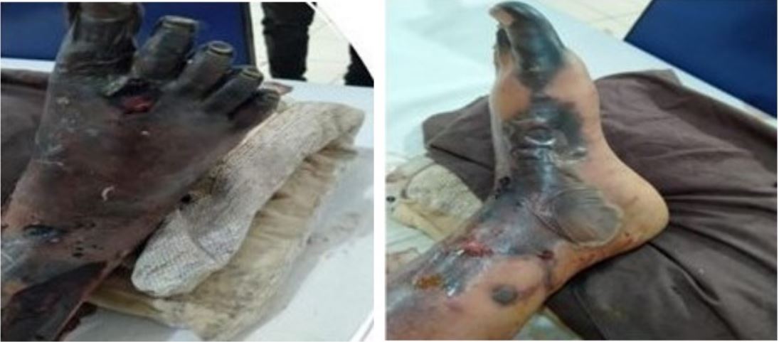
Table 1:Laboratory tests.
| Test Parameters | Observed Range | Reference Range |
|---|---|---|
|
Fibrinogen Degradation
Prod- uct, Plasma |
>5.0 ug/ml | <5 ug/ml |
| D-Dimer, Plasma | 25.2 mg/L FEU | <0.5 mg/L FEU |
| Anti-nuclear Antibodies (ANA) | Negative | - |
| Estimated Endpoint Titer | 1/80 | - |
|
Anti-Smooth muscle
Antibodies (ASMA) |
Negative | - |
|
Anti-mitochondrial
Antibodies (AMA) |
Negative | - |
| Hemoglobin | 7.3 g/dl | 11-14.5 g/dl |
| Hematocrit | 21.7% | 34.5-45.4 % |
| RBC | 2.33 x 10¹²/L | 3.61-5.2 x 10¹²/L |
| MCV | 93.1 fL | 78.1-95.3 fL |
| MCH | 31.3 pg | 25.3-31.7 pg |
| MCHC | 33.6 g/dl | 30.3-34.4 g/dl |
| RDW | 17.6% | 12.1-16.9 % |
| WBC | 15.2 x 10⁹/L | 4.6-10.8 x 10⁹/L |
| Neutrophils | 61.8% | 34.9-76.2% |
| Lymphocyte | 30.6% | 17.5-45% |
| Eosinophils | 0.4% | 0.3-7.4% |
| Monocyte | 6.7% | 3.9-10% |
| Basophils | 0.5% | 0-1% |
| Platelets | 97 x109/L | 154-433 x109/L |
| Serum B12 | 499 pg/ml | >201 pg/ml |
A broad range of infectious and noninfectious etiological factors are linked to symmetrical peripheral gangrene. The aetiology of SPG is heavily influenced by infection and the ensuing sepsis. Patients with SPG have been reported to develop sepsis from bacterial infections such as Pneumococcus, Neisseria meningitides, Staphylococcus aureus, and Streptococcus pyogenes [6]. Severe Plasmodium falciparum malaria infection is a rare condition that can lead to SPG [7]. Malignancy, hypovolemic shock, myeloproliferative disorders, vasospastic illnesses, connective tissue diseases such systemic lupus erythematosus (SLE), and antiphospholipid antibody syndrome are just a few examples of noninfectious causes of SPG [8]. Evidence also suggests drugs such as noradrenaline, adrenaline, and dopamine to be causative of the condition [6,7,9]. An underlying cause must be suspected, and timely diagnosis is necessary for SPG advancement [7].
Unfortunately, the precise pathophysiology of SPG is not fully known. Disseminated intravascular coagulation (DIC) at a low flow state is a component of the underlying mechanism. DIC was identified by Molos et al [6,11] as a significant underlying cause in 85% of patients who develop SPG [12]. Similar results were observed by Davis et al [13] in their group of 12 SPG patients. Immunosuppression, asplenism, hypothermia, diabetes mellitus, and renal insufficiency all negatively affect how SPG presents in individuals. SPG should be suspected when a patient exhibits cold extremities, pain in the distal extremities, cyanosis, and pallor. Before overt gangrene develops, early detection aids in stopping the course of ischemia alterations [14]. Ischemic alterations typically start at the edges and move closer. Peripheral vascular occlusive disease does not precede ischemic alterations. The big vessels’ distal pulses are still present. The microcirculation in the affected limbs may become occluded because of DIC and a related low-flow state, leading to ischemia and gangrene. According to a pathological analysis of specimens from amputated limbs, thrombi are primarily found in small blood arteries, sparing the larger ones. Additionally, there is no associated vasculitis [7]. This is significant because the underlying disease process must also be addressed; attention should not only be paid to the distal ischemia alterations [15].
Other causes of gangrene must be ruled out since symmetrical peripheral gangrene is a diagnosis of exclusion. Addressing and treating the underlying cause is the goal. Thomboangiitis obliterans, thromboembolic gangrene, calciphylaxis, and vasculitic gangrene are a few conditions similar to SPG. To rule out sepsis and DIC, an infection screen is crucial. Blood tests such as a complete blood count, blood culture, and peripheral smear aid in the early detection for sepsis [7]. Schistocytes on a peripheral smear could indicate DIC [6]. A peripheral smear will also aid in evaluating for cancer, and other SPG causes, such as Plasmodium falciparum. Patients can be evaluated for DIC by monitoring blood lactate levels, conducting a D-dimer assay, and assessing fibrin degradation products [7]. Blood tests for connective tissue disorders, such as the antinuclear antigen test (ANA) and antiphospholipid antibodies test (APLA) are additional helpful testing when warranted. Even when imaging tests like a Doppler USS can be performed, the management strategy remains unchanged. A peripheral Doppler examination will highlight the absence of involvement of the big peripheral vessels [7,11].
Patients with sepsis and DIC require sufficient resuscitation and broad-range antibiotics. Deficient clotting factors should be replaced to treat patients with a bleeding diathesis [7]. Treatment options for SPG patients include tissue plasminogen infusion and prostacyclin (epoprostenol) [6]. Leukapheresis, sympathetic blocking, and plasmapheresis may be helpful but only play a small part in SPG. It is debatable whether anticoagulants like heparin and aspirin should be used [6,16]. Streptokinaserin and streptokinase were not successful in stopping the progression of gangrene in SPG patients, according to Johansen and Hansen. Oral corticosteroids were also added, although they had little therapeutic benefit.
Most individuals with symmetrical peripheral gangrene invariably require an amputation, and mortality rates for the condition may reach 35% [17]. An initial nonsurgical approach is suitable, focusing on patient stabilization and underlying disease treatment. As a result, the gangrene can separate before an amputation is performed [11,12].
Conclusion
Therapeutic management of symmetrical peripheral gangrene is anecdotal and based on a few case series and case reports. No known treatment for SPG is consistently successful. The patient’s treatment needs to be customized. The underlying illness process must be addressed to have the greatest result for the patient while addressing patients with SPG. The best results come from managing the patient holistically and targeting the underlying pathophysiologic disease process rather than just concentrating on the ischemia and gangrenous processes in the peripheral limbs.
Declarations
Acknowledgments: The authors are very appreciative to the patient for the opportunity to learn as well as thankful to the hospital for providing support for completing this report.
Declaration of conflicting interests: The author(s) declared no potential conflicts of interest with respect to the research, authorship, and/or publication of this article.
Funding: The author (s) received no financial support for the research, authorship, and/or publication of this article.
Ethical approval: Our institution does not require ethical approval for reporting individual cases or case series.
Provenance and peer review Not: commissioned, externally peer reviewed.
Patient consent: Written informed consent from the patient’s legally authorized representative (LAR) for the publication of this case report.
References
- Acosta H, Forcada P, Bonjorn M, Mustafa A, Pilares P, Colomina J. Symmetrical Peripheral Gangrene: Report of Three Cases. Case Rep Crit Care. 2022; 2022: 8615420. doi: 10.1155/2022/8615420. PMID: 36278033; PMCID: PMC9581682.
- Davis M. P., Byrd J., Lior T., Rooke T. W. Symmetrical peripheral gangrene due to disseminated intravascular coagulation. Archives of Dermatology. 2001; 137(2): 139-140.
- Albano MN, Brazão SG, Caroço TV, Louro JM, Coelho MI, Costa Almeida CE, Reis LS, Costa Almeida CM. Rare case of symmetrical peripheral gangrene due to septic shock, disseminated intravascular coagulation and inotropic use. Ann Med Surg (Lond). 2018; 35: 103-107. doi: 10.1016/j.amsu.2018.09.025. PMID: 30294440; PMCID: PMC6168929.
- Batool W, Naveed S, Khan S, Ahmed SM. Postpartum septic shock presenting as symmetrical peripheral gangrene: A rare entity. Pak J Med Sci. 2023; 39(3): 916-918. doi: 10.12669/pjms.39.3.7277. PMID: 37250569; PMCID: PMC10214782.
- Khan MM, Shahid S, Zafar A, Khan NN, Bhatty S. Unusual Initial Presentation Of Primary Antiphospholipid Syndrome As Postpartum Symmetrical Peripheral Gangrene. J Ayub Med Coll Abbottabad. 2022; 34(1): 200-202. doi: 10.55519/JAMC-01-8695. PMID: 35466654.
- Sharma BD, Kabra SR, Gupta B. Symmetrical peripheral gangrene. Trop Doct. 2004; 34(1): 2‐4.
- Ghosh S, Bandyopadhyay D. Symmetrical peripheral gangrene. Indian J Dermatol Venereol Leprol. 2011; 77(2): 244‐248.
- Anuradha S, Prabhash K, Shome D, et al. Symmetric peripheral gangrene and falciparum malaria–an interesting association. J Assoc Physicians India. 1999; 47(7): 733-735.
- Deb SR, Kabir A, Khanum T, et al. Digital symmetrical peripheral gangrene: a rare male presentation of antiphospholipid anti body syndrome. J Dhaka Med Coll. 2016; 24(2): 152-155.
- Hayes MA, Yau EH, Hinds CJ, Watson JD. Symmetrical peripheral gangrene: association with noradrenaline administration. Intensive Care Med. 1992; 18(7): 433-436.
- Ghosh S, Bandyopadhyay D, Ghosh A. Symmetrical peripheral gangrene: a prospective study of 14 consecutive cases in a tertiary‐care hospital in eastern India. J Eur Acad Dermatol Venereol. 2010; 24(2): 214-218.
- Molos MA, Hall JC. Symmetrical peripheral gangrene and disseminated intravascular coagulation. Arch Dermatol. 1985; 121(8): 1057-1061.
- Davis MDP, Dy KM, Nelson S. Presentation and outcome of purpura fulminans associated with peripheral gangrene in 12 patients at Mayo Clinic. J Am Acad Dermatol. 2007; 57(6): 944‐956.
- Tripathy S, Rath B. Symmetric peripheral gangrene: catch it early!. J Emerg Trauma Shock. 2010; 3(2): 189.
- Parmar MS. Symmetrical peripheral gangrene: a rare but dreadful complication of sepsis. Can Med Assoc J. 2002; 167(9): 1037-1038.
- Davis MD, Byrd J, Lior T, Rooke TW. Symmetrical peripheral gangrene due to disseminated intravascular coagulation. Arch Dermatol. 2001; 137(2): 139-140.
- Johansen K, Hansen ST. Symmetrical peripheral gangrene (purpura fulminans) complicating pneumococcal sepsis. Am J Surg. 1993; 165(5): 642-645.