
Journal of Clinical Images and Medical Case Reports
ISSN 2766-7820
Clinical Image - Open Access, Volume 5
Shingles without hutchinson sign developing herpes zoster ophthalmicus and preseptal cellulitis
Liang Wang1; Juan Serralles Allongo1,2*
1University of Miami Miller School of Medicine, Miami, Florida, 33136, USA.
2University of Miami Hospital, University of Miami Miller School of Medicine, Miami, Florida, 33136, USA.
*Corresponding Author : Juan Serralles Allongo
University of Miami Hospital, University of Miami
Miller School of Medicine, Miami, Florida, 33136,
USA.
Email: jxs2909@med.miami.edu
Received : Feb 22, 2024
Accepted : Mar 11, 2024
Published : Mar 18, 2024
Archived : www.jcimcr.org
Copyright : © Allongo JS (2024).
Keywords: Herpes zoster; Herpes zoster ophthalmicus; Hutchinson sign; Shingles.
Citation: Wang L, Allongo JS. Shingles without hutchinson sign developing herpes zoster ophthalmicus and preseptal cellulitis. J Clin Images Med Case Rep. 2024; 5(3): 2926.
Description
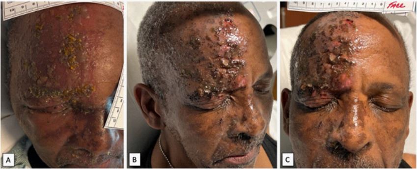
A previously healthy 69-year-old man presented to an emergency room with a vesicular rash on his right forehead in line with shingles (herpes zoster) secondary to varicella-zoster virus. The patient was negative for the Hutchinson sign, a rash involving the tip of the nose or nasal ridge, which indicates an increased risk of herpes zoster ophthalmicus (HZO). Only around 10-20% of shingles cases develop HZO, which is an emergent vision-threatening disease [1]. The patient reports history of chickenpox and denies any immunocompromising conditions [2]. He was sent home on a course of oral valaciclovir. Four days later he was brought to the emergency room with an enlarged, crusting rash encompassing the right side of his forehead to the eyelid with blurry vision and severe eyelid edema (Figure 1A). The rash was spread along the ophthalmic distribution of the trigeminal nerve completely within the V1 dermatome, indicating HZO. The ophthalmology exam showed chemosis, follicular conjunctivitis, and subconjunctival hemorrhages. There was no pain on extraocular movement or evidence of keratitis, uveitis, or retinitis. Laboratory tests were within normal limits, human immunodeficiency virus screening was non-reactive, and blood cultures showed no growth. A 14-day course of oral valaciclovir was started for varicella zoster. A seven-day course of vancomycin and ceftriaxone was started for potential orbital cellulitis. Bacitracin/polymyxin B ointment was applied to the eye and skin lesions to prevent bacterial superinfection. CT sinus later confirmed preseptal cellulitis with no ocular involvement. After 6 days of treatment, the patient reported no blurry vision and decreased pain with trace eyelid edema and healing of the skin with scabbing and fewer vesicles (Figure 1B and 1C). With the incidence of HZO almost tripling from 2004-2016 in the United States, it is increasingly important to vaccinate for shingles and monitor for HZO to prevent permanent vision loss [3].
Funding: None.
Conflict of interests: None.
References
- Zaal MJ, Völker-Dieben HJ, D’Amaro J. Prognostic value of Hutchinson’s sign in acute herpes zoster ophthalmicus. Graefes Arch Clin Exp Ophthalmol. 2003; 241(3): 187-91.
- Catron T, Hern HG. Herpes zoster ophthalmicus. West J Emerg Med. 2008; 9(3): 174-6.
- Voelker R. Increasing Cases of Shingles in the Eye Raise Key Questions. JAMA. 2019; 322(8): 712-714.