
Journal of Clinical Images and Medical Case Reports
ISSN 2766-7820
Case Report - Open Access, Volume 5
Innovative approach to traditional technique: A case of successfully fragmented giant gastrolithiasis by endoscopic injection method under super minimally invasive surgery
Jiafeng Wang; Fei Geng; Kang Du; Qianqian Chen*
Department of Gastroenterology, The First Medical Center of Chinese PLA General Hospital, Beijing, 100853, China.
*Corresponding Author : Qianqian Chen
Department of Gastroenterology, The First Medical Center of Chinese PLA General Hospital, Beijing, 100853, China.
Email: qian_qian_chen@163.com
Received : Mar 25, 2024
Accepted : Apr 12, 2024
Published : Apr 19, 2024
Archived : www.jcimcr.org
Copyright : © Chen Q (2024).
Abstract
Background: A case of massive gastrolithiasis was reported in which the drug treatment was not satisfactory. But it was successfully fragmented by endoscopic injection method.
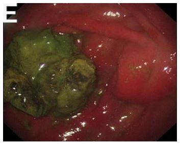
Case summary: We report the case of a 77-year-old man with presented to the abdominal distension after eating. There is a massive gastrolithiasis located at the site of the anastomosis of the residual stomach through gastroscopy. To get rid of gastrolithiasis, the hard outer surface of the rock was cracked open with a mouse tooth clamp snare to expose its soft inside. Then, by injecting sodium bicarbonate solution into the gastrolithiasis via an injection needle, the stone became loosen, and then the snare was used to break the stone into pieces.
Conclusion: The highly effective, short operation time, less pain, and no specific post-operative treatment can serve as the first-line treatment in the absence of adequate drug treatment. It can be marketed to community hospitals.
Keywords: Gastrolithiasis; Endoscopic injection; Sodium bicarbonate; Endoscopy; SMIS.
Core Tip: The clinical manifestations of gastrolithiasis lack specificity, and diagnosis usually requires gastroscopy, which usually have tendency to complications such as ulcers, bleeding, and intestinal obstruction. For giant plant gastrolithiasis, drug therapy is ineffective and surgical intervention may be necessary. We propose a technique for breaking up gastrolithiasis using existing tools, which is suitable for giant plant gastrolithiasis, can be performed even in community hospitals, and allows for rapid recovery of patients without the need for surgical intervention.
Citation: Wang J, Geng F, Du K, Chen Q. Innovative approach to traditional technique: A case of successfully fragmented giant gastrolithiasis by endoscopic injection method under super minimally invasive surgery. J Clin Images Med Case Rep. 2024; 5(4): 2999.
Introduction
The occurrence of gastrolithiasis is when there is food, matter, or foreign matter which is indigestible, unable to cross the pylorus in time, remains in the belly, or has congealed with the stomach mucus to form a solid mass. Gastrolithiasis may be classified into four groups based on its composition: plant gastrolithiasis, animal gastrolithiasis, drug gastrolithiasis, and mixed gastrolithiasis. Patients with gastrolithiasis don’t often have particular clinical symptoms, and ulcers, bleeding, and intestinal obstruction can make the condition worse [1,3]. At present, various therapies are used according to the composition and size of the gastrolithiasis. In the case of large plant gastrolithiasis, the therapeutic efficacy of the medicine is not effective, the efficacy of endoscopy is low, and even surgery is necessary [4]. With the increasing demand for improved quality of life among patients and the growing popularity of the concept of super minimally invasive surgery, we have developed a stone-breaking technology that utilizes existing tools for giant gastrolithiasis. It is possible to carry out this technology with no surgery, even at a primary health care facility, and enables quick recovery of patients.
Case presentation
Chief complaints: A 77-year-old Chinese man presented abdominal distension after eating persisted for over two months.
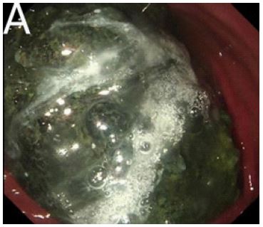
History of present illness: The patient occured abdominal distension and aggravated symptoms of acid reflux and heartburn after consuming a large amount of persimmons in March 2023. A follow-up gastroscopy was performed at a local hospital and found that there was a gastrolithiasis of 4 × 4 cm in residual gastric cavity.The therapeutic efficacy was unsatisfactory following a period of over 2 months of cola consumption, but improved to the size of 6 × 8 cm after a course of oral sodium bicarbonate tablets exceeding 1 month.
History of past illness: On June 18th, 2021, the patient underwent a Bilroth I gastrectomy due to adenocarcinoma of the gastric antrum.
Personal and family history: The patient denied any family history.
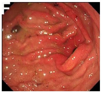
Diagnosis: Combined with the patient’s clinical manifestation and gastroscopy, the final diagnosis was gastrolithiasis.
Treatment
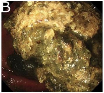
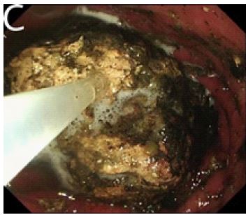
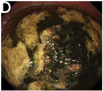
Innovative Approach to traditional technique used for fragmentation of giant gastrolithiasis by endoscopic injection method. The surface of the gastrolithiasis was encased in a rigid black shell, rendering it impervious to pharmacological intervention. The mouse tooth clamp snare was utilized to disrupt the rigid exterior of the gastrolithiasis and reveal its pliable interior. Sodium hydrogen carbonate solution is then inserted through an injection needle into the gastrolithiasis to release the stone inside. Finally, the snare was employed to fragment the stone into smaller pieces, facilitating its expulsion from the stomach.
Discussion
The highlight of this paper is to apply a new application of traditional methods to treat giant gastrolithiasis, which combined the advantages of currently widely used endoscopic lithotripsy techniques. The most frequent form of stomach gastrolithiasis is phytobezoars, which is more prevalent in North China [5]. According to prior literature, gastrolithiasis-inducing foods include hawthorn, persimmon, black dates, and glutinous rice balls. Rich in astringent tannins, pectin, and cellulose, they consumed on an empty stomach, react with proteins in the food to generate insoluble tannin-protein complexes under the influence of gastric acid. These complexes subsequently precipitate in the stomach and cling to indigestible plant fibers, such as persimmon fibers, culminating in the formation of rigid gastrolithiasis [6]. Gastrolithiasis is commonly observed in patients with autonomous nervous system disorders or those who have had previous stomach resection [7,8].
At present, treatment options for gastrolithiasis include drug dissolution therapy, endoscopic fragmentation, and surgical excision [9,10]. But their general condition is usually bad, and they do not tolerate the operation to remove the stones, or they will need to undergo an operation. In terms of medical treatment, commonly used methods include the use of solvents such as Coca-Cola, sodium bicarbonate, and enzymes for gastrolithiasis dissolution [10]. This medication has the ability to neutralize gastric acid and react with tannic acid, resulting in the formation of sodium tannate. This compound is characterized by its brittleness and susceptibility to breakdown into carbondioxide and water. The carbondioxide bubbles generated during this process facilitate the disintegration of gastric gastrolithiasis, rendering them fragile and readily dissolvable [11]. For patients suffering from giant gastrolithiasis, prolonged formation time of gastrolithiasis, and unsuccessful drug litholysis, endoscopic lithotripsy is the recommended course of action. The conventional methods for endoscopic treatment of large plant gastrolithiasis involve the use of foreign body forceps, snares, and baskets for stone retrieval, as well as injection needles and self-made snares. Although foreign body forceps are widely used and easy to operate, repeated friction between the endoscope and the pharynx during the procedure can cause damage to the pharyngeal mucosa, leading to secondary pharyngitis. Long surgeries can exacerbate pain, and conventional treatment methods often necessitate multiple endoscopic procedures, resulting in high treatment costs. Moreover, currently available snares on the market have a limited diameter and cannot fully encircle the stone for cutting, rendering them susceptible to deformation during subsequent cutting processes. Similarly, stone retrieval baskets also encounter similar issues. Although injection needles can accurately dissolve gastrolithiasis, penetrating the surface of gastrolithiasis with a hard shell remains challenging. The newly developed self-made snare boasts an adjustable diameter, a high success rate in a single attempt, a short surgery time, and reduces the risk of damage to the patient’s throat and esophagus by eliminating the need for repeated removal [12]. However, the assimilation and mastery of novel devices requires a significant investment of time.
Innovative approach for traditional technique was used. Gastrolithiasis that respond poorly to drug therapy is usually coated with a tough outer layer, which prevents the entry of medicines. In order to solve this problem, we first use a mouse-tooth forceps trap to remove the surface hard shell, exposing the relatively loose inside layer. Then, we inject the sodium bicarbonate precisely into the stone using a needle, which rapidly dissolving the stone. This is an effective treatment measure that allows drugs to enter the interior and loosen the stone. Subsequently, a snare is used to break the stone into pieces, allowing it to be expelled from the stomach, achieving the concept of rapid healing and full recovery. We achieved successful treatment of the patient with conventional instruments and medication, obviating the necessity for specialized devices or the acquisition of novel surgical techniques, thereby facilitating expeditious application. Because of its short operation period, rapid recovery, and low incidence of complications, the necessity of specialist post-surgery care made it an excellent method of distribution to primary care facilities.
Conclusion
The highly effective, short operation time, less pain, and no specific post-operative treatment can serve as the first-line treatment in the absence of adequate drug treatment. It can be marketed to community hospitals.
Declarations
Author contributions: Wang JF, and Geng F contributed to manuscript writing and editing; Du K contributed to follow-up; Chen QQ contributed to conceptualization and supervision; all authors have read and approved the final manuscript.
Grant support: National Key Research & Development Program of China (No. 2022YFC2503600).
Acknowledgements: We declare that we have no financial and personal relationships with other people or organizations that can inappropriately influence our work.
References
- Meng Y, Luo YB, Zhou YH et al. Characteristics of clinical and endoscopic findings of phytobezoars: analysis of 77 cases(in Chinese).Journal of Gastroenterology and Hepatology. 2022; 31: 784-787.
- Sun H, Gao C, Wei X. Gastrolithiasis with incomplete intestinal obstruction in the perioperative period of percutaneous transluminal coronary intervention: one case report. Ann Palliat Med. 2020; 9: 4389-4393. doi:10.21037/apm-20-467.
- Akrami M, Sasani MR. Dietary Habits Affect Quality of Life: Bowel Obstruction Caused by Phytobezoar. Iran J Public Health. 2016; 45: 1080-1082.
- Paschos KA, Chatzigeorgiadis A. Pathophysiological and clinical aspects of the diagnosis and treatment of bezoars. Ann Gastroenterol. 2019; 32: 224-232. doi:10.20524/aog.2019.0370.
- Fan CQ, Zhang PB, Yu J et al. Effect of Sequential Gastroscopy and 5% Sodium Bicarbonate Solution on Gastrolith (in Chinese). Chinese Journal of Digestive endoscopy. 2013; 30: 404-406.
- Liu LN, Wang L, Jia SJ et al. Clinical Features, Risk Factors, and Endoscopic Treatment of Bezoars: A Retrospective Analysis from a Single Center in Northern China. Med Sci Monit. 2020; 26: 926539. doi:10.12659/msm.926539.
- Hemmasi G, Zanganeh E, Hosseini SA et al. Risk factors, endoscopic findings, and treatments in upper gastrointestinal bezoars: multi-center experience in Iran. Gastroenterol Hepatol Bed Bench. 2021; 14: 160-164.
- Liu Y, Li M, Jin L et al. Upper Gastrointestinal Obstruction Caused by Gastrolithiasis After Laparoscopic Roux-en-Y Gastric Bypass: a Case Report. Obes Surg. 2019; 29: 1937-1938. doi:10.1007/s11695-018-03674-7.
- Ren XF, Ai YW, Zhao F et al. Application of painless electronic gastroscopy in treatment of gastric concretion (in Chinese). World Journal of Chinese Digestion. 2014; 22: 4816-4819.
- Zhang LY, Cui Z, Ji FZ et al. Retrospective analysis of efficiency and expense of fragmentation of gastric bezoar through endoscopy in different period (in Chinese). Chinese journal of Endoscopy. 2015; 21: 875-877.
- Dong HQ, Li GP, Wang HL et al. (in Chinese). X-ray diagnosis andaerogenic anenttherapy of crataegus gastrolith: a report of 196 cases.Journal of Practical Medical Imaging. 2003; 329-330.
- Xu W, Liu XB, Li SB et al. Self-made wire loop snare successfully treats gastric persimmon stone under endoscopy. World J Clin Cases. 2022; 10: 6428-6436. doi: 10.12998/wjcc.v10.i19.6428.