
Journal of Clinical Images and Medical Case Reports
ISSN 2766-7820
Clinical Image - Open Access, Volume 5
Stevens-Johnson syndrome and toxic epidermal necrolysis: Vulvar and vaginal manifestations
Karamaroudis Stefanos1*; Karamperis Alexandros1; Alexiou Nikolaos1; Soumakis Konstantinos1; Sotiropoulou-Karamperi Georgia2
1Department of Obstetrics and Gynecology, General Hospital of Elefsis “Thriassio”, Elefsis, Attiki, Greece.
2Medical Center, Nea Makri, 1st Health Authority of Attica, Greece.
*Corresponding Author : Karamaroudis Stefanosa
Department of Obstetrics and Gynecology, General Hospital of Elefsis “Thriassio”, Elefsis, Attiki, Greece.
Email: stefanos.karamaroudis@gmail.com
Received : Apr 15, 2024
Accepted : May 01, 2024
Published : May 08, 2024
Archived : www.jcimcr.org
Copyright : © Stefanos K (2024).
Keywords: Stevens-Johnson syndrome; Toxic epidermal necrolysis; Vulvovaginal epidermal necrolysis.
Abbreviations: SJS: Stevens-Johnson Syndrome; TEN: Toxic Epidermal Necrolysis; PPI: Proton Pump Inhibitor; BSA: Body Surface Area.
Citation: Stefanos K, Alexandros K, Nikolaos A, Konstantinos S, Sotiropoulou-Karamperi G. Stevens-Johnson syndrome and toxic epidermal necrolysis: Vulvar and vaginal manifestations. J Clin Images Med Case Rep. 2024; 5(5): 3037.
Description
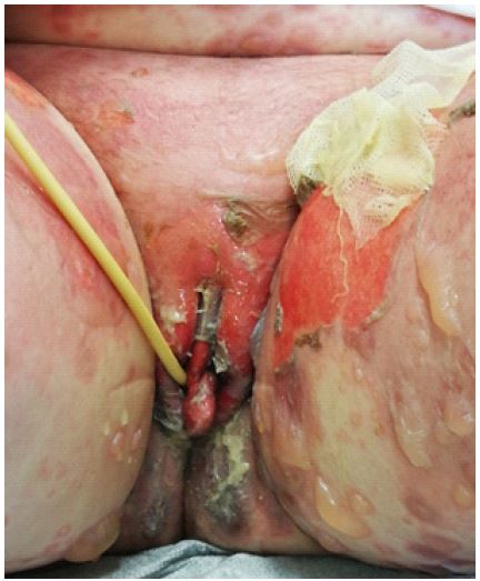
A 75-year-old woman was admitted to our emergency department due to SJS and TEN to investigate the involvement of the vulva and the vaginal mucosa and make a consultation. The woman’s medical history included hypertension, managed with a beta-blocker and a combination hydrochlorothiazide and valsartan, and gastro-esophageal reflux disease managed with a PPI which recently started.
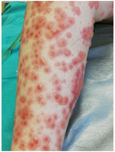
SJS and TEN are severe mucocutaneous adverse reactions characterized by epidermal necrosis. Both are variants of the same disease process and categorized by the percentage of BSA with epidermal detachment [1]:
- SJS: < 10% of BSA
- SJS/TEN: 10-30%
- TEN: >30%
There is no certain cause but many factors have been suspected such as infections or medications like non-steroidal anti-inflammatory drugs (e.g. oxicams family etc.), anti-neoplatic agents or antiepileptics. Women have higher risk to develop the disease. HLA-B15 and HLA-A31 phenotypes seem to be related with increased risk.
Treatment and proper care of the involved tissues is required to minimize the long-term effects of the disease. About 70% with SJS or/and TEN have vulvovaginal involvement and up to one third of the treated patients will have chronic complications [3]. There is no consensus on the optimal treatment. Usually a combination of topical agents such as corticosteroids and estrogens, vaginal dilator therapy and Foley catheter to avoid agglutination and menstrual supression is the preferred management. The role of the gynecologist is to determine the vaginal mucosa involvement and give proper guidelines as mentioned above [2]. Most of the times speculum examination can be carried out only under anesthesia. Finally, the primary care gynecologist should not forget that any non-traumatic erosion or ulcer of the vagina or vulva might be the initial presentation of the syndrome. In our case we can only suspect that the recently started PPI was the trigger factor. The patient diascharged after long hospitalization in the Burn Unit of our hospital [5].
Declarations
Acknowledgments: We would like to thank the patient for allowing us to share this clinical image.
Disclosure: The authors declare no potential conflict of interest.
References
- Crowder CA, Jeney SES, Kraus CN, Bernal N, Lane F. Vulvovaginal involvement in Stevens-Johnson syndrome and toxic epidermal necrolysis: management and techniques used to reduce gynecologic sequelae. Int J Dermatol. 2022; 61(2): 158-163. doi:10.1111/ijd.15676.
- DenAdel MA, Hendrickson SE, Fuchs E. Stevens Johnson Syndrome: Past, Present, and Future Directions Gynecologic Manifestations and Management in SJS/TEN. Front Med (Lausanne). 2022; 9: 874445. Published 2022 Jul 4. doi:10.3389/fmed.2022.874445.
- Hollingsworth J, Park SU, Bhagavathi V, Green A, Philips N. Stevens-Johnson syndrome with vulvar involvement: A case report and literature review. Case Rep Womens Health. 2022; 34: 00404. Published 2022 Mar 14. doi: 10.1016/j.crwh.2022. 00404.
- Mergler R, Chuang M. Stevens Johnson Syndrome with Vaginal Pain and Lesions as Initial Presentation. Am J Case Rep. 2018; 19: 1519-1521. Published 2018 Dec 21. doi:10.12659/AJCR.912123.
- Daldoul M, Charfi O, Bouattour E, et al. Pantoprazole-induced Stevens-Johnson Syndrome: A Case-report. Curr Drug Saf. 2024; 19(1): 148-150. doi: 10.2174/1574886318666230224092818.