
Journal of Clinical Images and Medical Case Reports
ISSN 2766-7820
Short Report - Open Access, Volume 5
Maxillary psammomatoid juvenile ossifying fibroma: A rare clinicopathological entity
Vera Eiró1*; Madalena Ferreira2; Cláudia Mendes1; Tatiana Gigante1; Sofia Moura Antunes2; Júlio Matias1
1Plastic Reconstructive Surgery, Centro Hospitalar Lisboa Ocidental, Lisbon, Portugal.
2Pediatrics Department, Hospital Dr. José de Almeida, Lisbon, Portugal.
*Corresponding Author : Vera Eiró
Plastic Reconstructive Surgery, Centro Hospitalar Lisboa Ocidental, Lisbon, Portugal.
Email: vera.s.eiro@gmail.com
Received : May 14, 2024
Accepted : Jun 14, 2024
Published : Jun 21, 2024
Archived : www.jcimcr.org
Copyright : © Eiró V (2024).
Abstract
The juvenile psammomatoid ossifying fibroma (JPOF) is a rare benign fibroosseous neoplasm of the craniofacial bones. It has predilection for sinusal locations, in young patients, with aggressive behavior and high recurrence rate. Surgical intervention is needed to secure a complete resection of the neoplasm. We present one unusual case of a young man presenting with a manifestation of JPOF in the maxilla, an unusual location, that was approached and completely removed via Caldwell-Luc approach. Early detection avoided exaggerated distortion of the anatomy, allowing an excision without complex reconstruction needs, and no recurrences in the postoperative follow-up.
Keywords: Juvenile ossifying fibroma; Craniofacial; Caldwell-Luc; Case report.
Citation: Eiró V, Ferreira M, Mendes C, Gigante T, Antunes SM et.al. Maxillary psammomatoid juvenile ossifying fibroma: A rare clinicopathological entity. J Clin Images Med Case Rep. 2024; 5(6): 3135.
Introduction
Juvenile psammomatoid ossifying fibroma (JPOF) is a rare benign, although aggressive, fibro-osseous neoplasm of the craniofacial bones. It is a subtype of juvenile ossifying fibroma with particular characteristics, since it is most commonly seen in early ages, with most cases reported in young teenage years, and has no gender predominance. The most common location is the paranasal sinus [1] with less common locations identified in the jaws (mandible and maxilla) and temporal bone, with male predominance in these locations and a more aggressive behaviour [2,3]. The predilection for certain locations distinguishes from the trabecular type that predominantly affects the jaws [4]. On CT the findings are consistent, a well delimitated mass with a thick wall of bone density, but variable density and multiloculated appearance inside and calcified areas. These imagiological findings are important in the jaws, to differentiate from fibrous dysplasia – without well-demarcated borders –, that has a limited clinical course [3]. Surgical intervention is needed to secure a complete resection of the neoplasm, with preservation of as much anatomical structures as possible, especially in younger ages when bone growth is a concern. The aggressive growth and tendency to recur is age related, and long follow-up periods must be considered, although there is no standard protocol [5]. The aim of this article is to report a clinical case of one unusual case of JPOF in the maxilla.
Case presentation
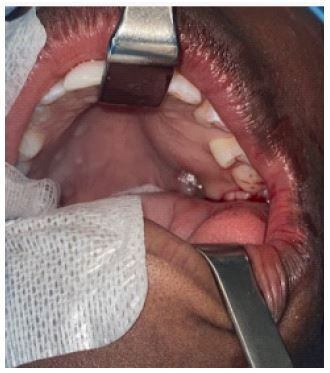
We report a case of a sixteen-year-old boy presenting to the emergency room with a painless swelling in the left maxillary area just near the pyramidal aperture, with continuous growth reported in the previous three months. No relevant family and medical history. There was no history of toothache, tooth treatment or discharge of purulent content. At clinical exam there was a mild but visible distortion of the left nasolabial fold and at examination of the oral cavity a hard palate cortical expansion was identifiable (Figure 1).
A facial CT (Figure 2) scan was requested, showing an apparently round cystic lesion, well defined, with 3.3 x 3 cm, in the second quadrant motivating a gap between 2.4 and 2.5 teeth, remodeling the floor of the nasal cavity was found as well as obliteration of the maxillary sinus. A few calcifications were noticeable inside of the mass. The diagnostic hypothesis of odontogenic keratocystic tumor, myxoma and ameloblastoma were also contemplated.


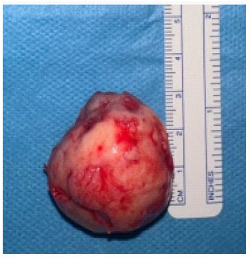
Considering its volume and continued rapid growth, the surgical excision was programmed. In the operating room, under general anesthesia, a vestibular approach was performed with external approach (Caldwell-Luc technique) and removal of what proved to be a solid, rich in connective tissue, mass. Curettage, and removal of an impacted tooth in the sinus were also executed (Figure 3). During the intervention, the cortical continuity was preserved, and no bone reconstruction was needed. The surgery and postoperative period were uneventful, and the patient was discharged on the second postoperative day.
There were no complications during the recovery period. The specimen (Figure 4) was sent for histopathologic analysis and the concluded diagnostic was juvenile psammomatoid ossifying fibroma. At nine months of follow up, there were no recurrences or new lesions.
Conclusion
Although it is a benign pathology with no description of malignant transformation in literature, its aggressiveness and expansile behavior can interfere with function (obstructive airway, visual changes), bacterial infections (recurrent sinusitis or associated with intracranial extension [1]) and psychosocial interactions (visible deformity). Therefore, the treatment of choice is early surgical excision, before the remodeling of anatomic structures nearby dictates a need for a more complex reconstruction, with higher morbidity associated. Special care must be taken to completely remove the entire lesion, to avoid recurrences. If so, it is associated with very good prognosis. Notes The authors have declared that no competing interests exist.
References
- Peterson, Brandon R., and Brenda L. Nelson. “Juvenile Active Ossifying Fibroma.” Head and Neck Pathology. 2014; 9(93,7): 384-386. link.springer.com/article/10.1007/s12105-014-0595-8, https://doi.org/10.1007/s12105-014- 0595-8 2 Tolentino, Elen S., et al. “Psammomatoid Juvenile Ossifying Fibroma: An Analysis of.
- Cases Affecting the Mandible with Review of the Literature.” Oral Surgery, Oral Medicine, Oral Pathology and Oral Radiology. 2012; 113(6): 40-45. www.sciencedirect.com/science/article/pii/S2212440311006742, https://doi.org/10.1016/j.oooo.2011.08.005.
- Hee Han, Moon, et al. “Sinonasal Psammomatoid Ossifying Fibromas: CT and MR Manifestations.” AJNR - American Journal of Neuroradiology. 1991; 12: 25-30.
- El-Mofty, Samir. “Psammomatoid and Trabecular Juvenile Ossifying Fibroma of the Craniofacial Skeleton: Two Distinct Clinicopathologic Entities.” Oral Surgery, Oral Medicine, Oral Pathology, Oral Radiology, and Endodontology. 2002; 93(3): 296-304. https://doi.org/10.1067/moe.2002.121545.
- Sarode, Sachin C, et al. “Juvenile Psammomatoid Ossifying Fibroma: A Review.” Oral Oncology. 47(12): 2011; 1110-1116. www.sciencedirect.com/science/article/pii/S1368837511007548, https://doi.org/10.1016/j.oraloncology.2011.06.513