
Journal of Clinical Images and Medical Case Reports
ISSN 2766-7820
Clinical Image - Open Access, Volume 5
Lumbar disk herniation mimicking a spinal tumor: A case report
Jihane EL Houssni*; Asaad EL Bakkari; Nada Adjou; Sanae Jellal; Youssef Omor; Rachida Latib; Sanae Amalik
National Institute of Oncology Rabat, University Mohammed V of Rabat, Morocco.
*Corresponding Author : Jihane EL Houssni
National Institute of Oncology Rabat, University Mohammed V of Rabat, Morocco.
Email: elhoussnijihane@gmail.com
Received : Jun 13, 2024
Accepted : Jun 27, 2024
Published : Jul 04, 2024
Archived : www.jcimcr.org
Copyright : © Houssni JE (2024).
Keywords: Lumbar disk herniation; MRI; Differential diagnosis.
Citation: Houssni JE, Bakkari AE, Adjou N, Jellal S, Omor Y, et al. Lumbar disk herniation mimicking a spinal tumor: A case report. J Clin Images Med Case Rep. 2024; 5(7): 3153.
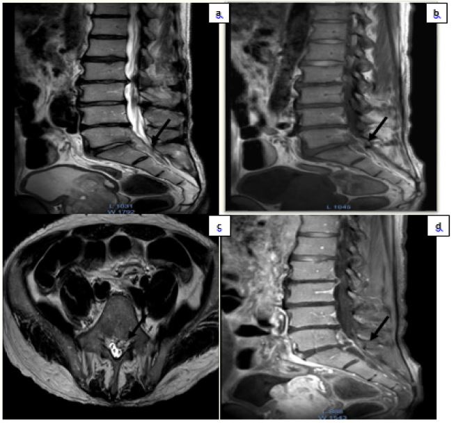
Description
Patient aged 54 years, operated on 4 months ago for gastric adenocarcinoma, currently undergoing adjuvant chemotherapy, presenting with paresis of the lower limbs. An MRI of the lumbar spine was performed, revealing a sequestrated extra-dural mass adjacent to the vertebral bodies of L5, S1, and S2, with a median, para-median, and left foraminal position, reducing the anterior epidural space, with an ascending and descending trajectory showing T1-weighted hypointensity and T2-weighted hyperintensity. This mass conflicts with the emergence of the left S1 and the left L5 nerve root, showing peripheral enhancement after gadolinium injection, and extending over 66 mm. It is associated with a large sequestrated migratory L5-S1 disc herniation, conflicting with the left L5 and S1 nerve roots.
Diagnosing a large lumbar disc herniation can be challenging, as a severe disc herniation often mimics spinal tumors [1]. The clinical presentation is difficult to distinguish from other causes of lumbar canal stenosis, such as synovial cysts, epidural hematomas, metastases, and tumors [1]. Sequestrated disc herniations generally appear as heterogeneously hypo- to isointense on T1- and T2-weighted MRI images [2]. MRI with gadolinium injection is useful for differentiating a disc herniation from tumors and other epidural lesions, as the disc fragment typically shows peripheral enhancement. A herniated disc fragment rarely presents central enhancement, which is secondary to vascular granulation tissue infiltrating the fragment, but it is never associated with enhancement of the spinal meninges, a characteristic of neoplastic lesions such as lymphoma and neurofibroma [2]. An epidural abscess is often located in the posterior epidural space, appearing as low or moderate signal on T1-weighted images and high signal on T2-weighted images with homogeneous or peripheral enhancement [2]. Neurilemmomas are often located in the peridural space; they are isointense on T1-weighted images, hyperintense on T2-weighted images, and enhanced after gadolinium injection [2]. Meningiomas are more common in the thoracic spine and are often located within the dura mater. They are isointense on T1- and T2-weighted images and enhanced on contrast MRI [3].
In our case, the mass showed T2 isointensity and T1 hypointensity with peripheral enhancement, typical of a disc fragment. Nevertheless, the larger size and elongated shape of the lesion caused confusion, as lumbar disc herniations are usually much smaller and round. Another factor of confusion was the fact that this longitudinal mass was related to two different intervertebral spaces with slight reduction in their height on MRI [2].
In conclusion, sequestrated lumbar disc fragments must be considered in the differential diagnosis of extra-dural masses of the spinal canal even when their shape and large size are counterintuitive [2].
Declarations
Conflict of interests: No conflicts of interest.
Funding statement: All authors have no funding source to declare.
Ethical approval: Not required.
Consent: Written consent has been obtained.
References
- Chu EC, Lin A, Huang KHK, Cheung G, Lee WT. A Severe Disc Herniation Mimics Spinal Tumor. Cureus. 2023; 15(3): 36545. doi: 10.7759/cureus.36545. PMID: 36968683; PMCID: PMC10033246.
- Dimogerontas G, Paidakakos NA, Konstantinidis E. Voluminous free disk fragment mimicking an extradural tumor. Neurol Med Chir (Tokyo). 2012; 52(9): 656-8. doi: 10.2176/nmc.52.656. PMID: 23006881.
- Li ST, Zhang T, Shi XW, Liu H, Yang CW, Zhen P, Li SK. Lumbar disc sequestration mimicking a tumor: Report of four cases and a literature review. World J Clin Cases. 2022; 10(9): 2883-2894. doi: 10.12998/wjcc.v10.i9.2883. PMID: 35434096; PMCID: PMC8968809.