
Journal of Clinical Images and Medical Case Reports
ISSN 2766-7820
Short Report - Open Access, Volume 5
Neonatal cytosteatonecrosis: Hypercalcemia with nephrocalcinosis
Hani S*; El Qadiry R; Lagrine M; Nassih H; Bourahouat A; Ait Sab I
Department of Pediatrics B, University Hospital Centre Mohamed VI, Marrakech, Morocco.
*Corresponding Author : S Hani
Department of Pediatrics B, University Hospital Centre Mohamed VI, Marrakech, Morocco.
Email: dr.sorayahani@gmail.com
Received : Jun 14, 2024
Accepted : Jun 28, 2024
Published : Jul 05, 2024
Archived : www.jcimcr.org
Copyright : © Hani S (2024).
Abstract
Cytosteonecrosis (CSN) is a rare pathology that is poorly described in the medical literature. Its pathogenesis is still poorly elucidated, and it generally develops in the neonatal period. Diagnosed clinically, it presents as indurated, purplish skin patches on fair skin or hyperchromic patches on black skin, often localized on the face, trunk, buttocks and roots of limbs. Histologically, it is a lobular panniculitis, with fat necrosis and infi ltrate granulomatous cells (lymphocytes, histiocytes, fi broblasts and giant cells) and calcium deposits. The course of the disease is spontaneously favorable within a few weeks, with no sequelae. However, in some cases, the onset of major hypercalcemia can lead to life-threatening complications. We report a case of neonatal cytosteatonecrosis complicated by hypercalcemia and nephrocalcinosis.
Keywords: Cytosteatonecrosis; Neonatal; Hypercalcemia.
Citation: Hani S, El Qadiry R, Lagrine M, Nassih H, Bourahouat A, et al. Neonatal cytosteatonecrosis: Hypercalcemia with nephrocalcinosis. J Clin Images Med Case Rep. 2024; 5(7): 3157.
Introduction
Cytosteonecrosis of the newborn is an acute hypodermatitis that develops during the first few days of life. It takes the form of indurated, purplish skin patches on fair skin or hyperchromic patches on black skin, often localized on the face, trunk, buttocks and roots of limbs. Histologically, the normal epidermis and dermis are underlain by lobular panniculitis with foci of eosinophilic necrosis of adipose tissue encompassing optically empty intra-adipocytic radial clefts, corresponding to lipid dissolution and crystallization [1]. The main risk situations classically reported are fetal macrosomia, often in the context of a diabetic mother, perinatal asphyxia, severe hypothermia and tissue trauma during instrumental maneuvers or neonatal resuscitation [2]. The course of cytosteonecrosis is usually benign. However, in certain cases, the onset of severe hypercalcemia can be responsible for complications that can be life-threatening [3]. We report a case of neonatal cytosteatonecrosis complicated by hypercalcemia and nephrocalcinosis.
Clinical observations
We report the case of a 48-day-old infant admitted to pediatric ward B for late postprandial vomiting, from an unrelated marriage to a 32-year-old mother, vaginal delivery, with notion of perinatal asphyxia classified as stage I according to Sarnat’s classification.
At 1 day of age, the mother noticed induration on both cheeks, which she considered normal. She consulted 6 weeks later for postprandial vomiting in a context of apyrexia, and on admission, diffuse subcutaneous indurations were noted on the 2 lower limbs, trunk and cheeks, with no signs of dehydration. Biological findings included hypercalcemia at 160 mg/l (normal calcemia: 95-102 mg/l), hypertriglyceridemia at 2.40 with normal lipid balance, thrombocytosis at 752000 (platelet count: 150,000-400,000), phosphorus levels and renal function.
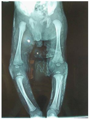
Bone calcifications
Renal ultrasound revealed stage III nephrocalcinosis and ETF was normal. Management consisted of hyperhydration with infusion of 0.9% saline at 100 ml/kg/d, forced diuresis with furosemide 1 mg/kg/d and low-dose corticosteroid therapy with prednisone 0.4 mg/kg/d. Daily clinical monitoring, with twice-daily monitoring of laboratory results. The evolution was marked by regression of the calcemia level until normalization at 108 after 6 days of treatment with total regression of lesions at 3 months of evolution, with a normal renal ultrasound at 4 months.
Discussion
NSC complicated by hypercalcemia and nephrocalcinosis is rarely described in the literature. Subcutaneous lesions generally appear within the first 4 weeks of life, but can occur earlier, in the first few days of life, as was the case in our observation. These lesions appear as subcutaneous concretions or indurated purplish-red plaques located mainly on the limbs, trunk and face [4,5]. The topography of the lesions observed in our patient is in line with those reported in the literature.
Several hypotheses have been put forward in relation to a fatty tissue anomaly: disturbed fat metabolism with excess saturated fats in subcutaneous tissue, hypoxia due to neonatal suffering or hypothermia favoring saturated fat crystallization and fat necrosis [6]. This pathogenesis explains the risk factors generally incriminated: perinatal asphyxia, obstetrical trauma, resuscitation with inappropriate maneuvers, abdominal compressions [2]. Other factors have been implicated, notably therapeutic hypothermia, which has become widely indicated in the treatment of severe perinatal asphyxia [1]. Other maternal factors such as gestational diabetes, pre-eclampsia, placenta previa, cocaine or calcium antagonists, and Rh incompatibility have been reported [7].
The only concern with neonatal NSC is the occurrence of severe hypercalcemia. This hypercalcemia can manifest itself as feeding difficulties, vomiting, anorexia and agitation [8-11]. It frequently occurs in disseminated forms of NSC, as was the case with our patient. The mechanism of onset of hypercalcemia is unclear, and several mechanisms may be involved:
Necrosis of adipose cells leading to an increase in prostaglandins with activation of osteoclasts; release of calcium by necrotic adipocytes; abnormal production of 1,25-dihydroxyvitamin-D by macrophages increasing bone turnover.
The risk of hypercalcemia correlates directly with the extent of skin lesions. Major hypercalcemia carries the risk of tissue deposits, in particular nephrocalcinosis as in our observation. Deposits in the heart (inter-atrial septum, valves), liver and inferior vena cava have also been described [11]. As soon as the diagnosis is made, blood calcium levels must be monitored [4], and a search for calcium deposits in the kidneys, liver and other viscera should be the rule. Malignant or life-threatening hypercalcemia requires intensive care combining hyperhydration with isotonic sodium solution, Henle’s loop diuretics and short-term corticosteroid therapy, starting with a low dose of prednisone (0.4 mg/kg per day) [12]. Abnormal bone calcium release can also be blocked by calcitonin injections (4 IU/kg intramuscular or subcutaneous), which have a rapid (12 to 24 hours) but modest effect, and are usually of short duration (four to six hours) and well tolerated in children. This conventional treatment (in addition to a low-calcium diet and abstention from vitamin-D administration) is not always sufficient to normalize blood calcium levels, and sometimes necessitates recourse to the use of biphosphonates first described for the treatment of hypercalcemia associated with cytosteatonecrosis by Rice and Rivkees. These biphosphonates inhibit bone resorption through their action on osteoclasts. Their use in paediatrics is hampered by fears of potential side-effects on bone growth. They are well tolerated both intravenously and per os. Among biphosphonates, there are several molecules, and dosage in this indication is poorly codifi ed. The low frequency of this condition and the age of the children preclude pharmacological studies [13]. In our observation, saline hyperhydration, furosemide and prednisone were sufficient to normalize blood calcium levels without visceral sequelae.
Conclusion
NSC is a rare entity that deserves to be recognized. It should not be regarded as a benign disease, as hypercalcemia, a classic complication, can be life-threatening. Within two months of the diagnosis of NSC, weekly monitoring of ionized calcium should be performed. As soon as hypercalcemia occurs, a low-calcium diet is required, sometimes accompanied by hyperhydration and the use of diuretics or low-dose corticosteroids. In severe forms that do not respond to treatment, biphosphonates can provide a therapeutic alternative that is not without risk.
References
- Bégon E, Blum L, Petit jean B, Jacomet L, Merbouche S, Moguelet P, et al. Neonatal subcutaneous adiponecrosis (cytosteatonecrosis) and hypercalcemia after therapeutic hypothermia. Ann Dermatol Venereol. 2012; 139(8-9): 601-2.
- Rachid Abilkassem1 & Nezha Dini1, Mohamed Oukabli2, Mohamed Kmari1, Aomar Agadr1 Association of neonatal cytosteatonecrosis, hypertriglyceridemia and hypercalcemia: about an observation Pan African medical journal Published. 2012.
- Barbier C, Cneude F, Deliège R, El Kohen R, Kremp O, Leclerc F. Cytostéatonécrose néonatale: attention à l’hypercalcémie sévère. Arch Pediatr. 2003; 10(8): 713-5.
- Tizki S, Lehlimi M, Habzi A, Benomar S. Neonatal cytosteatonecrosis: beware of hypercalcemia, even late! Journal de Pédiatrie et de Puériculture. 2013; 26(2): 105-8.
- Dudink J, Walther FJ, Beekman RP. Subcutaneous fat necrosis of the newborn: hypercalcaemia with hepatic and atrial myocardial Calcification. Arch Dis Child Fetal Neonatal. 2003; 88(4): 343-5.
- Perrotta R, Virzi D, Tarico MS. A rare case of congenital ulcerated subcutaneous fat necrosis of the newborn. J Plast Reconstr Aesth Surg. 2010; 63(11): 801-2.
- Germanaud D, Hadj-Rabia S, Parsy C, Abadie V. Neonatal cytosteatonecrosis complicated by symptomatic hypercalcemia: efficacy of low-dose corticosteroids. Arch Pediatr. 2007; 14(2): 167-9.
- Barltrop D. Hypercalcaemia associated with neonatal subcutaneous fat necrosis. Arch Dis Child. 1963; 38(201): 516-8.
- Singalavanija S, Limponsanurak W, Wannaprasert T. Subcutaneous fat necrosis of the newborn. J Med Assoc Thai. 2007; 90(6): 1214-20.
- Lewis HM, Ferryman S, Gatrad AR, Moss C. Subcutaneous fatnecrosis of the newborn associated with hypercalcae. Soc Med. 1994; 87(8): 482-483.
- Mahé E, De Prost Y. Cytosteatonecrosis of the newborn Ann Dermatol Venereol. 2007; 134(5): 494-8.
- Karochristou K, Siahanidou T, Kakourou-Tsivitanidou T, Stefa-naki K, Mandyla H. Subcutaneous fat necrosis and hypocalcaemia. J Perinatol. 2006; 26: 64-66
- Tran JT, Sheth AP. Complications of subcutaneous fat necrosis of the newborn: a case report and review of the literature. Pediatr Dermatol. 2003; 20(3): 257-61.