
Journal of Clinical Images and Medical Case Reports
ISSN 2766-7820
Case Report - Open Access, Volume 5
Didelphys uterus: A case report and review of the literature
Yafet Abebe*; Gebresilassie Andualem
Department of Obstetrics and Gynecology, Myungsung Christian Medical Center, Addis Ababa, Gerji, Ethiopia.
*Corresponding Author : Yafet Abebe
Department of Obstetrics and Gynecology, Myungsung Christian Medical Center, Addis Ababa, Gerji, Ethiopia.
Email: yafet.abebe5@gmail.com
Received : Jun 15, 2024
Accepted : Jul 04, 2024
Published : Jul 11, 2024
Archived : www.jcimcr.org
Copyright : © Abebe Y (2024).
Abstract
Abnormal embryological development of the Mullerian ducts results Mullerian duct anomalies, which are congenital defects of the female genital system. The female genital tract and the urinary tract are closely related anatomically and embryologically and it has been reported that about 10% of infants are born with some genitourinary system abnormality. One of the rarest types of MDAs is a didelphys uterus, sometimes referred to as a “double uterus”. This report discusses a case of didelphys uterus that successfully conceived, carried her pregnancy to term, and delivered without any significant complications. Patient is a 29-year-old primigravida from Addis Ababa, who had uncomplicated antenatal care from 33+1 weeks. Patient presented with the onset of labour to MCM Hospital at 38+2 weeks. On per vaginal examination a non-communicating, a thick vaginal septum extending from the introit to the cervix, and two cervixes were found. Patient had a caesarean delivery with intra-operative findings of didelphylic uterus each bearing single tube and ovary, gravid left hemi uterus. The surgery was uneventful and EBL 700 mls.
Citation: Abebe Y, Andualem G. Didelphys uterus: A case report and review of the literature. J Clin Images Med Case Rep. 2024; 5(7): 3164.
Introduction
Obstetricians and gynaecologists often come with some of the more intriguing illnesses that are related to malformations of the Muller-duct system. The primordial anlage of the female reproductive system is the Muller-ducts. When this mechanism is interfered with, a variety of deformities can arise [1]. One of the rarest congenital anomalies of the female reproductive system is uterine didelphys, which arises when the para-mesonephric ducts’ tissues fail to unite or resorb completely between weeks 6 and 22 of gestation. This can lead to a variety of clinical manifestations, including a double uterus and mal developed vagina, such as a single or double vagina.
It is widely acknowledged that, when compared to a normal uterus, having a uterine abnormality is associated with worse pregnancy outcomes, including higher risks of spontaneous abortion, early labor, caesarean delivery owing to breech presentation, and fewer live infants. The severity of these effects, however, varies depending on the kind of uterine anomaly [2].
Term delivery rates for didelphys and uniconuate uteri are approximately 45%, but untreated bicornuate and septate uteri have poor pregnancy outcomes with term delivery rates of only approximately 40% [4,5,6]. With term delivery rates of roughly 65%, an arculate uterus is linked to a marginally improved but still compromised pregnancy outcome.
Case presentation
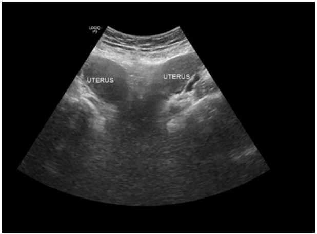
A 29 year-old primigravida woman at 33+1 weeks of gestation presented to the MCM hospital to establish her antenatal care [5]. Patient had uncomplicated antenatal care from 33+1 weeks and did not give any history of signs of threatened abortion or signs of threatened preterm labour. Her routine ANC investigations and vital signs remained within normal limits and fetal ultrasound was also normal. Foetus showed appropriate growth. Pregnancy was spontaneously conceived and planned, diagnosed with a urine pregnancy test after two missed periods. She had taken 2 doses of tetanus toxoid (TT) vaccine and was taking prenatal vitamins. The course of her antenatal visit in the index pregnancy was uneventful.
At 38+2 week gestation, the patient presented to MCM hospital with pushing down pain of 11 hours duration. On physical examination, she was a young woman, healthy looking, afebrile, not pale, well hydrated, no pedal edema. Her pulse rate was 84 beats per minutes, full volume, and regular. Blood pressure was 110/70 mmHg. Respiratory rate was 20 cycle per minute. On per abdomen examination, abdomen was uniformly enlarged, term sized gravid uterus, singleton fetus was palpable in longitudinal lie, cephalic presentation, fetal heart rate was 140 beats per minutes. On per vaginal examination a non-communicating, a thick vaginal septum extending from the introit to the cervix, and two cervixes, normal urethral and anal orifices. The patient did not report having dyspareunia, dysmenorrhea or chronic abdominal pain in the past. So she was admitted to labour ward to follow the progress of labor. The patient preferred a caesarean section delivery so the patient underwent emergency lower segment caesarean section under spinal anaesthesia in view of CDMRs.
Diagnostic assessment
The full blood count shows RBC of 4.42 x 10⁹/L, a white blood cell count of 15 x 109/L, platelet count of 163,000/ml. Blood group was A positive. Antibodies to HIV and VDRL were non-reactive. HbsAg was negative. Urinalysis was essentially normal.


Therapeutic interventions
Informed consent was obtained for Emergency caesarean section, and she was reviewed by the anaesthetists. After Spinal Anaesthesia was done, she underwent caesarean section. She delivered at 38+2 weeks with the intra-operative findings of didelphylic uterus each bearing single tube and ovary, gravid left hemi uterus. The right hemi uterus was about the size of an adult fist. Baby delivered by vertex presentation. The outcome was an alive male neonate weighing 2.4 kg with Apgar score of 8 and 9 in the 1 and 5 mins, placenta was anterior and fundal. The uterus was closed in double layers. Abdomen was closed in layers. The surgery was uneventful and EBL 700 mls. There were no renal anomalies on subsequent USG. She did well post operatively and was discharged home on the second post op day after counselling for family planning.
Follow up and outcome of interventions
she was seen at the post-natal clinic 1 week after her discharge with no complaints.
Patient perspective: She was satisfied with the quality of care she received during her antenatal and post-natal visits.
Consent: Informed consent was obtained from the patient for images and information provided in this case report?
Discussion
Comparing a didelphys uterus to other anomalies included in the Buttram and Gibbons classification, a didelphys uterus is still an extremely uncommon Mullerian duct phenomenon. The majority of information about this uterine anomaly’s clinical importance and outcomes is derived from small retrospective, observational, or case studies.
The very low occurrence of the anomaly in the population and the fact that more research has been focused on the more prevalent malformations-arcuate, septate, and bicornuate as well as the varied outcomes of these studies can be attributed to these factors.0
While dyspareunia or dysmenorrhea may be present in women with didelphys uterus in the presence of a thick, occasionally blocking vaginal septum, most women with this condition are asymptomatic. In addition to causing hematocolpos or hematometrocolpos, this blocked vaginal septum can also cause chronic abdominal pain. Rarely, reports of endometriosis and genital neoplasms are linked to didelphys uterus instances.
When anatomical anomalies were observed that required additional investigation, a conventional speculum exam typically resulted in an initial suspicion of the disorder and a diagnosis [1,7]. Furthermore, kidney abnormalities may be found alongside uterine abnormalities because Mullerian ducts often originate in conjunction with Wolffian ducts.
In a prospective observational study of the reproductive outcome of women with various uterine anomalies compared to a normal uterus, Acién et al discovered that the rate of term delivery for a didelphys uterus was significantly lower than the normal uterus group, but not as low as the bicornuate and septate groups [7].
A didelphys uterus has been shown in many case reports to occur as a part of a syndrome, more specifically called, Herlyn-Werner-Wunderlich (HWW) syndrome, also known as obstructed hemivagina and ipsilateral renal anomaly (OHVIRA) [6]. It is a very rare congenital anomaly of the urogenital tract involving Mullerian ducts and Wolffian
Structures, and it is characterized by the triad of didelphys uterus, obstructed hemivagina, and ipsilateral renal agenesis [8].
Conclusion
In conclusion, women who have uterine anomalies that are congenital, like uterine didelphys, can be diagnosed with a high degree of suspicion. Management of this patient can pose a serious challenge to the obstetrician, especially as pregnancy in this group of patients are associated with adverse outcomes. However, individualized management of patients with uterine didelphys and delivery by elective caesarean section produces favorable pregnancy outcome. Overall, there is a scarcity of literature on the didelphys uterus at the moment. As a result, further research is needed to better understand the reproductive and gestational outcomes so that doctors may provide appropriate advice and treatment to their patients.
References
- Heinonen PK, Saarikoski S, Pystynen P. Reproductive performance of women with uterine anomalies. An evaluation of 182 cases. Acta Obstet Gynecol Scand. 1982; 61: 157-62.
- Stanislav S, Stoyan K, Angel Y. Pregnancy and childbirth in Uterine Didelphys: A Report of Three Cases. Medicina (Kaunas). 2020; 56(4): 198. PubMed | Google Scholar.
- Omeed P, Dana B, Caria P, Wendy W. Uterine Didelphys in a Pregnant Mother. Open Journal of Obstetrics and Gynecology. 2018; 8(13).
- Raga F, Bauset C, Remohi J, Bonilla-Musoles F, Simón C, Pellicer A. Reproductive impact of congenital Müllerian anomalies. Human Reproduction. 1997; 12(10): 2277-2281.
- Grimbizis GF, Camus M, Tarlatzis BC, Bontis JN, Devroey P. Clinical implications of uterine malformations and hysteroscopic treatment results. Human Reproduction Update. 2001; 7(2): 161-174.
- Acién P. Reproductive performance of women with uterine malformations. Human Reproduction. 1993; 8(1): 122-126.
- Heinonen PK. Clinical implications of the didelphic uterus: long-term follow-up of 49 cases. European Journal of Obstetrics & Gynecology and Reproductive Biology. 2000; 91(2): 183-190.
- Stanislavsky A. Herlyn-Werner-Wunderlich syndrome (HWW). Radiopaedia. 2015.