Journal of Clinical Images and Medical Case Reports

ISSN 2766-7820
Case Report - Open Access, Volume 5

Gastric outlet obstruction secondary to giant traumatic pancreatic pseudocyst

Muhammad Zakwan Bin Yahya1*; Kew Thean Yean2; Malinda Abd Majid3; Tan Tiong Keat4

1International Islamic University of Malaysia (IIUM), Malaysia.

2Hospital Censalor Tunku Mukhriz (HCTM), Malaysia.

3Hospital Kuala Lumpur (HKL), Malaysia.

4Hospital Sultan Abdul Halim, Sungai Petani, Malaysia.

*Corresponding Author : Zakwan Bin Yahya M
International Islamic University of Malaysia (IIUM), Malaysia.
Email: [email protected]

Received : Sep 13, 2024

Accepted : Oct 09, 2024

Published : Oct 16, 2024

Archived : www.jcimcr.org

Copyright : © Yahya MZB (2024).

Abstract

Traumatic pancreatic injury is rare due to its retroperitoneal location and relative protection to the organ. Pancreatic pseudocyst formation may occur following pancreatic trauma, but giant pseudocyst rarely develops. Early management is recommended for giant pancreatic pseudocysts before clinical deterioration. We presented a case report with gastric outlet obstruction symptoms in the casualty department. Imaging investigations Computed Tomography (CT) confirmed the presence of pancreatic transection with giant pseudocyst formation. He underwent endoscopic cystogastrostomy with immediate resolution of the symptoms. Imaging is essential for earlier detection and severity of the pancreatic injury and its complication like pancreatic pseudocyst formation. Imaging follows up are needed to monitor disease recurrence or suspected pancreatic duct injury and assess pancreatic ductal integrity before transluminal stent removal.

Keywords: Pancreatic pseudocyst; Gastric outlet obstruction; Pancreatic transection.

Citation: Yahya MZB, Yean KT, Majid MA, Keat TT. Gastric outlet obstruction secondary to giant traumatic pancreatic pseudocyst. J Clin Images Med Case Rep. 2024; 5(10): 3298.

Introduction

Pancreatic injuries are uncommon due to their retroperitoneal location, which protects the organ. It accounts for less than 2% of blunt abdominal trauma injuries [1]. It is associated with significant morbidity and mortality, with a mortality rate as high as 34% [2]. Common blunt traumatic pancreatic injury complications include traumatic pancreatitis, pseudocyst formation and fistula, and less commonly causing gastrointestinal bleeding and splenic vein thrombosis [1]. Traumatic transection resulting in complete pancreatic rupture commonly occurs in the line of the superior mesenteric vein at the neck of the gland [3]. The reported incidence of pancreatic pseudocyst following trauma varies and may reach 30% of pancreatic trauma cases [4]. Pancreatic pseudocysts are defined as fluid collection rich with pancreatic enzymes like amylase and surrounded by fibrous tissue wall rather than the true epithelial lining. The pseudocyst has direct or indirect communication with the ductal system, which is caused by pancreatic ductal disruption following inflammation, trauma or obstruction [5]. Giant pancreatic pseudocysts are rare, where the major diameter of the cyst measures 10 cm and above [6]. Local compression onto the duodenum can cause gastric outlet obstruction, where early satiety, nausea, vomiting and weight loss are manifestations of this complication [7].

We reported a 33-year-old male with a giant pancreatic pseudocyst following blunt abdominal trauma, which presented with gastric outlet obstruction symptoms as the initial presentation.

Figure 1: Sagittal reconstructed images of the CECT abdomen show irregular non-enhancing hypodensity with fluid attenuation seen at the pancreatic body (white arrows). Anterolaterally, it is connected to a large well-defined hypodense rim enhancing collection (line arrow) measuring approximately 10.3 cm x 12.8 cm x 23.4 cm (APxWxCC) with an attenuation value of 6-12HU.

Figure 2: Axial image of CECT abdomen show mass effect onto surrounding structures. Abrupt tapering of the calibre of proximal D3 segment of duodenum (arrow)with dilatation of duodenal bulb and D2 segment of duodenum (line arrow) in reconstructed coronal image. On further history taking, he was allegedly assaulted and stepped on the abdomen repeatedly by three people at a caretaker centre two weeks prior. The patient was referred to the Hepatobiliary team in a tertiary centre for further management. He underwent endoscopic cystogastrostomy. Scope findings showed that the posterior wall of the stomach has a prominent bulge from the pseudocyst. Cystogastrostomy was applied to the posterior wall of the stomach, and the pseudocyst was cannulated. Double pigtail deployed x2 (10Fr 10 cm and 10Fr 5 cm). The flow of pseudocyst content into the stomach after drainage. The post-operative impression was pseudocyst post-pancreatic trauma. Repeated CECT abdomen a month after the procedure on 6th July 2021.

Figure 3: Shows presence of two double pigtail catheters with one end is seen anterior to the previously seen communication of the large pseudocyst with the hypodense pancreatic body with another end seen within the pylorus. h. shows resolution of the large pancreatic pseudocyst.

Case presentation

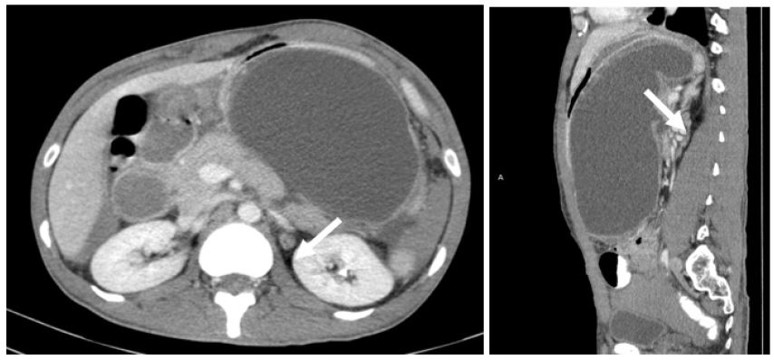
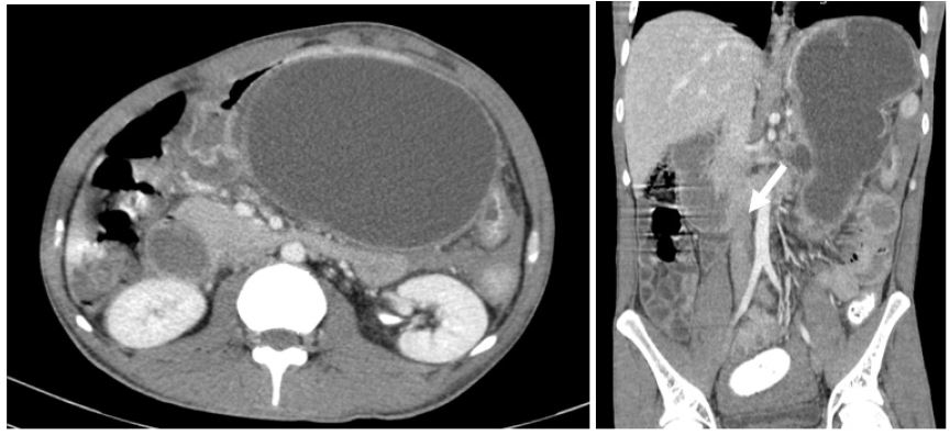
A 33-year-old male with underlying schizophrenia presented to a district hospital with a complaint of persistent vomiting for the past two weeks, three to four times a day, primarily fluid content. Associated with diarrhoea, abdominal pain, lethargy and unable to tolerate orally for two weeks. Clinical examination revelaed mass over left-sided abdomen extending to the right paraumbilical region, firm in consistency. Otherwise, the vital signs are all normal. Serum amylase was elevated 386 U/L (normal value: 40-140 U/L). Contrast-Enhanced Computed Tomography (CECT) abdomen and pelvis were performed and shows pancreatic transection with pseudocyst causing gastric outlet obstruction.

Table 1: American Association for the Surgery of Trauma – Organ Injury Scale (ASST - OIS) [1].
Grade Injury Description of the pancreatic injury
I Hematoma Minor contusion without ductal injury
Laceration Superficial laceration without ductal injury
II Hematoma Major contusion without ductal injury or tissue loss
Laceration Major laceration without ductal injury or tissue loss
III Laceration Distal transection or pancreatic parenchymal injury with ductal injury
IV Laceration Proximal transection or pancreatic parenchymal injury involving the ampulla
V Laceration Massive disruption of the pancreatic head

Discussion

Pancreatic injury is rare but can cause dangerous complications. It resulted from direct compression of the gland against the lumbar vertebrae [3]. Slim young males and children are at higher risk of injury due to a a lack of a protective retroperitoneal fat pad [2]. Pancreatic parenchymal destruction or complete rupture of the gland usually results from direct compression of the gland against the lumbar vertebrae [3]. The reported occurrence of pseudocyst following pancreatic trauma varies and may reach up to 30% [4]. The pathogenesis is believed to be due to disruption of the main pancreatic duct or peripheral ductules, causing leakage and activation of the pancreatic enzyme [7]. Two third of patients with pseudocysts have demonstrable communications between the cyst and the pancreatic duct, while the rest are not demonstrable, likely due to a sealed connection resulting from an inflammatory reaction [5]. Patients with pseudocyst do not have specific symptoms; however, it should be suspected in a patient presented with persistent abdominal pain, abdominal mass, anorexia or unable to tolerate orally after an episode of pancreatitis or abdominal trauma [5]. There are a few classifications and grading for pancreatic trauma, and the most widely used is the Organ Injury Scale (OIS) developed by the American Association For The Surgery Of Trauma (ASST) [1]. The main determinants of the scoring are the location of the injury and the presence of parenchymal or ductal injury (Table 1).

Elevation of serum amylase is neither sensitive nor specific for diagnosing pancreatic injury. However, hyperamylasemia is a sign of probable pancreatic injury because elevated serum amylase has been observed in 75% of patients with blunt abdominal trauma and proven pancreatic injury [8]. Serum amylase has limited utility in diagnosing pancreatic pseudocyst, which is often elevated, but maybe within normal ranges [5]. Imaging is pivotal in the early detection of pancreatic injury and its related complications like pseudocyst formation for early management and treatment plan. CT has a sensitivity and specificity of as high as 80% for detecting pancreatic injury. However, CT may underestimate the severity of the damage, and normal initial findings may not exclude pancreatic injury [8].

In 2020, a systematic review suggested that Endoscopic Ultrasound (EUS), Endoscopic Retrograde Pancreatography (ERCP) and Magnetic Resonance Cholangiopancreatography (MRCP) with- or without secretin are all accurate in diagnosing any suspected disruption or disconnection of the pancreatic duct with a sensitivity of 100% each [9]. Being non-invasive, faster and more readily available than ERCP, MRCP has appeared as the preferred choice for direct imaging of the pancreatic duct. MRCP may also demonstrate abnormalities not visible at ERCP, such as fluid collections upstream of the site of duct transection and helpful in assessing parenchymal injury [10]. Ultrasound and CT scans are good imaging tools to confirm the diagnosis of pancreatic pseudocyst, as in our case. Considering the operator dependence of ultrasound, the diagnostic sensitivity and specificity are still high, with percentages of 88-100% and 92-98% each. CT scan has the highest sensitivity (82-100%) and specificity (98%) [11]. In addition, CT scan delivers extra detailed information regarding the surrounding anatomy and can demonstrate additional pathology, including pancreatic duct dilatation and calcification or extension of the pseudocyst outside the lesser sac. Life-threatening complications are reported in 10% of pseudocysts cases and include infection, bleeding from ruptured pseudoaneurysm in the wall of the pseudocyst and pseudocyst-peritoneal fistula causing pancreatic ascites, gastric or rarely oesophagal variceal bleed due to splenic or portal vein thrombosis, and erosion of the pseudocyst into the portal venous system and arteries [12]. Asymptomatic pseudocysts measuring up to 6 cm in diameter can be safely managed conservatively and monitored with serial imaging [6]. Symptomatic pseudocyst or complications like infected pseudocyst, gastric outlet obstruction, haemorrhage or biliary obstruction are the main indications for intervention [5]. In our case, the patient requires intervention due to the large size of the pseudocyst with gastric outlet obstruction symptoms. Treatment options for pancreatic pseudocysts include open surgery, percutaneous drainage, and laparoscopic or endoscopic cystogastrostomy [13]. In 2013, a randomized trial comparing the efficacy of endoscopic and surgical cystogastrostomy for pancreatic pseudocyst drainage showed that both methods were of similar efficacy. However, endoscopic treatment was associated with shorter hospital stays and patients’ better physical and mental health at a lower cost [6]. Kean et al. concluded that endoscopic drainage has superior rates of treatment success, lower rate of re-intervention and shorter length of hospital stay than percutaneous drainage [14]. According to an expert consensus in 2020, endoscopic transluminal drainage was the experts’ first preferred treatment strategy for patients with infected (peri) pancreatic necrosis and a confirmed disrupted or disconnected pancreatic duct. Most experts indicated removing transluminal stent only after evaluation of pancreatic duct integrity on imaging [15]. Our patient underwent an endoscopic cystogastrostomy; however, the long hospital stay was due to COVID-19 infection. Otherwise, pertaining to the pancreatic pseudocyst, he showed clinical and imaging improvement following the endoscopic cystogastrostomy.

Conclusion

Traumatic pancreatic injury is rare due to its retroperitoneal location with relative protection to the organ. Pancreatic pseudocyst formation is a late complication following pancreatic trauma; however, giant pseudocysts rarely develop. Imaging (CT scan and MRCP) plays an important role for earlier detection and assessing the severity of pancreatic injury as well as its complications like pancreatic pseudocyst formation. Early treatment is recommended for giant pancreatic pseudocysts before clinical deterioration. Imaging follow up is needed to monitor recurrence or suspected pancreatic duct injury and assess the pancreatic ductal integrity prior to transluminal stent removal.

References

  1. Ull C, Bensch S, Schildhauer TA, Swol J. Blunt Pancreatic Injury in Major Trauma: Decision-Making between Nonoperative and Operative Treatment. Case Rep Surg. 2018; 2018: 6197261.
  2. Duggan W, Hannan E, Brosnan C, O’Sullivan S, Conlon K. Conservative management of complete traumatic pancreatic body transection; A case report. Int J Surg Case Rep. 2020; 71: 222-4.
  3. Olah A, Issekutz A, Haulik L, Makay R. Pancreatic transection from blunt abdominal trauma: Early versus delayed diagnosis and surgical management. Dig Surg. 2003; 20(5): 408-14.
  4. Lin BC, Fang JF, Wong YC, Liu NJ. Blunt pancreatic trauma and pseudocyst: management of major pancreatic duct injury. Injury. 2007; 38(5): 588-93.
  5. Habashi S, Draganov PV. Pancreatic pseudocyst. World J Gastroenterol. 2009; 15(1): 38-47.
  6. Udeshika WAE, Herath H, Dassanayake SUB, Pahalagamage SP, Kulatunga A. A case report of giant pancreatic pseudocyst following acute pancreatitis: Experience with endoscopic internal drainage. BMC Res Notes. 2018; 11(1): 262.
  7. S Sahu, S Raghuvanshi, S Agarwal, D Bahl, Sachan P. Gastric Outlet Obstruction As Initial Presentation of Traumatic Pancreatic Pseudocyst. The Internet Journal of Surgery. 2006; 12(1).
  8. Ankouz A, Elbouhadouti H, Lamrani J, Taleb KA, Louchi A. Pancreatic transection due to blunt trauma. J Emerg Trauma Shock. 2010; 3(1): 76-8.
  9. Timmerhuis HC, Van Dijk SM, Verdonk RC, Bollen TL, Bruno MJ, et al. Various Modalities Accurate in Diagnosing a Disrupted or Disconnected Pancreatic Duct in Acute Pancreatitis: A Systematic Review. Dig Dis Sci. 2021; 66(5): 1415-24.
  10. Debi U, Kaur R, Prasad KK, Sinha SK, Sinha A, et al. Pancreatic trauma: A concise review. World J Gastroenterol. 2013; 19(47): 9003-11.
  11. Setiawati Dea Alberta, Suwardi, Saadhi Ikhdin, Santoso PG. Traumatic Giant Pancreatic Pseudocyst in Pediatric Patient A Rare Case Report. Asian Journal of Case Reports in Surgery. 2021.
  12. Ghulam Rasul SI, Mohamad IS, Zakaria Z, Syed Aziz SH. Pancreatic Pseudocyst with Gastric Outlet Obstruction: A Case Report and Literature Review. IOSR Journal of Dental and Medical Sciences. 2014; 13(2): 56-8.
  13. Gull-Zareen Khan Sial, Abid Quddus Qazi, Yusuf MA. Endoscopic Cystogastrostomy- Minimally Invasive Approach for Psueodocyst. Association of Pediatric Surgeons of Pakistan (APSP). 2015.
  14. Keane MG, Sze SF, Cieplik N, Murray S, Johnson GJ, et al. Endoscopic versus percutaneous drainage of symptomatic pancreatic fluid collections: A 14-year experience from a tertiary hepatobiliary centre. Surg Endosc. 2016; 30(9): 3730-40.
  15. Boxhoorn L, Timmerhuis HC, Verdonk RC, Besselink MG, Bollen TL, et al. Diagnosis and treatment of pancreatic duct disruption or disconnection: An international expert survey and case vignette study. HPB (Oxford). 2021.