Journal of Clinical Images and Medical Case Reports

ISSN 2766-7820
Research Article - Open Access, Volume 5

Prevalence of tooth transpositions and associated dental anomalies: A CBCT study

Kübra Çam1*; A Zeynep Zengin2

1Research Assistant, Department of Oral and Maxillofacial Radiology, Faculty of Dentistry, Ondokuzmayıs University, Samsun, Turkey.

2Associate Professor, Department of Oral and Maxillofacial Radiology, Faculty of Dentistry, Ondokuzmayıs University, Samsun, Turkey.

*Corresponding Author : Kübra Çam
Research Assistant, Department of Oral and Maxillofacial Radiology, Faculty of Dentistry, Ondokuzmayıs University, Samsun, Turkey. Tel: 0362-312-19-19;
Email: [email protected]

Received : Sep 19, 2024

Accepted : Oct 22, 2024

Published : Oct 29, 2024

Archived : www.jcimcr.org

Copyright : © Çam K (2024).

Abstract

Purpose: The aim of this study is to evaluate the frequency and type of dental transposition and its relationship with other dental anomalies and pathologies using Cone Beam Computed Tomography (CBCT).

Materials methods: Images of patients who had CBCT taken for various reasons between 2012 and 2023 were evaluated for the presence of dental transposition. The demographic data of the patients, the presence of transposed teeth, their number, location, unilateral or bilateral nature, dental transposition classifications defined in the literature, other adjacent dental anomalies and pathologies were examined.

Results: CBCT images of 5000 patients were examined and 42 transposed teeth were found in 39 of the patients (0.78%). It was seen unilaterally in 92.3% of cases and bilaterally in 7.7% of cases. The most common was maxillary canine-first premolar transposition (47.61%), and the least common was maxillary central-lateral transposition (2.38%). The presence of persistent primary teeth was the highest in the relevant region (61.53%). External root resorption of tooth roots adjacent to transposed teeth was diagnosed in 17.94% of cases.

Conclusion: The prevalence of transposition in Turkish society was found to be 0.78%. This study is the first to examine dental transpositions using CBCT in the Turkish population. External root resorption was observed in a number of the adjacent teeth showing transposition. Detailed CBCT examination is very important in diagnosing malocclusion and other problems that may occur due to transposed teeth.

Keywords: Transposition; Prevalence; CBCT.

Citation: Çam K, Zengin ZA. Prevalence of tooth transpositions and associated dental anomalies: A CBCT study. J Clin Images Med Case Rep. 2024; 5(10): 3316.

Introduction

Transposition is a rare dental anomaly that typically involves the positional change of two adjacent teeth [1]. Displacement of the crowns and roots of the related teeth in the dental arch is defined as complete transposition, and the displacement of crowns while the roots remain in their normal position is defined as partial transposition [2]. Although its prevalence is reported to be approximately 0.4% [3], it has not been reported in primary dentition [4].

Tooth transpositions are more common in the maxilla than in the mandible, and the most common transposed teeth are the maxillary permanent canines [5]. They are more common in women and they may be unilateral or bilateral [6]. However, there are studies in literature which report that they present most commonly unilateral and on the left side [7]. The aetiology of transposition is not fully known yet. Both genetic and environmental factors have a role [3]. Various factors can be listed as etiological factors, such as trauma, early loss of primary teeth, long term retention, delayed root resorption and positional displacement of developing tooth buds; however, the main etiological factors are genetic [4-6].

Six types of dental transpositions have been defined in literature [6,8].

These are;

Upper canine-first premolar transposition (Mx.C.P1) (Figure 1).

Upper canine -lateral incisor transposition (Mx.C.12).

Transposition of the upper canine to the first molar region (Mx.C to M1).

Upper lateral incisor-central incisor transposition (Mx.12.11).

Transposition of the upper canine to the central incisor region (Mx.C to 11) [6] (Figure 2).

Lower lateral incisor-canine transposition (Mn.12.C) [8].

Dental transposition is usually associated with other dental anomalies such as agenesis, primary canine retention and wedge-shaped lateral teeth [2,9]. They can also cause complications such as severe rotations of the teeth, malpositions, resorption or malformation of the adjacent tooth 1. The aim of this study is to evaluate the frequency and type of dental transposition and its relationship with other dental anomalies/pathologies by using CBCT.

Material and methods

ChordomaThe protocol and method of the study was approved by Clinical Research Ethics Committee of Ondokuz Mayıs University [OMU KAEK 2023-290].

Images of patients who had CBCT for various reasons between 2012 and 2023 were examined retrospectively in terms of the presence of dental transposition. The patients who had pathological conditions such as cysts, tumours and fractures in the area to be examined and the images which had inadequate diagnostic quality for various reasons were not included in the study. Demographic data of patients, presence of transposed teeth, number and location of transposed teeth, whether they were unilateral or bilateral, complete or partial, pathologies related with these teeth, other dental anomalies and their dental transposition classifications as described in the literature were examined.

Results

CBCT images of 5000 patients were examined retrospectively. A total of 42 dental transpositions were found in 39 of the patients (0.78%). 53.8% (n=21) of the cases were in female patients, while 46.2% (n=18) were in male patients. Mean age of female patients was found as 23.47 (14-70), while mean age of male patients was found as 18.33 (14-25).

Transposition was partial in 61.9% (n=26) of the transposed teeth, while it was complete in 38.1% (n=16). Transposition was unilateral in 92.3% of the patients (n=36), while it was bilateral in 7.7% (n=3) (Figure 3).

92.3% (n=36) of the teeth were in maxilla, while 7.7% (n=3) were in mandible. While 58.9% (n=23) of the transpositions in the maxilla were on the right, 41.1% (n=16) were on the left. Transposition was present only on the right in mandible.

While the most common type of transposition (Type A, Mx.C.P1) was between canine and first premolar teeth (47.6%). Maxillary central-lateral (Type D, Mx.12.11) was the least common type (2.4%). Type of transposition between maxillary canine-first molar tooth (Type C, Mx.C to M1), which was included in the classification, was not found in this study (Table 1).

In 30 (76.92%) patients, different dental anomalies were observed on the affected side. Most commonly, 27 persistent primary teeth were found in 24 (61.53%) patients in the relevant area (Figure 4). While 25 of the persistent primary teeth were primary canines, 1 was primary lateral and 1 was primary second molar tooth. Other than these, microdontia was found in 2 lateral teeth of 2 patients (5.12%), 2 impacted teeth (1 central and 1. premolar tooth) were found in 2 patients (5.12%), one supernumerary tooth was found in one patient (2.56%) and one congenitally missing lateral tooth (maxillary lateral) was found in 4 patients (10.25%).

External root resorption (n=7, 17.94%) was found in the adjacent tooth in 6 patients, and in the transposed tooth in one patient (Figure 5). Three resorptions were present in the lateral teeth, and three were in the central teeth, one resorption was first premolar tooth. Most of the teeth (71.42%) with resorption were included in lateral-canine transpositions (Type B, Mx.C.12).

Figure 1: Right maxillary canine- first premolar tooth transposition-Type A (Mx.C.P1) CBCT panoramic section image (Red arrow: Transposed right maxillary canine, Black arrow: Transposed right maxillary first premolar tooth).

Figure 2: Left maxillary canine - lateral tooth transposition - Type B (Mx.C.12) CBCT panoramic section image (Black arrow: Transposed left maxillary canine, Red arrow: Transposed left maxillary lateral tooth).

Figure 3: Bilateral canine-premolar tooth transposition-Type A (Mx.C.P1) CBCT axial section image (Black arrows: Bilateral transposed maxillary canines, Red arrows: Bilateral transposed maxillary first premolars).

Figure 4: Left maxillary canine -lateral tooth transposition -Type B (Mx.C.12) CBCT axial section image (red arrow; left maxillary canine tooth, black arrow; left upper lateral tooth, black arrow head; persistent primary canine tooth in the relevant area).

Figure 5: Right maxillary canine - lateral tooth transposition - Type B (Mx.C.12). External root resorption present in the right maxillary lateral tooth on the CBCT tangential section image (black arrow).

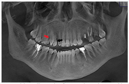
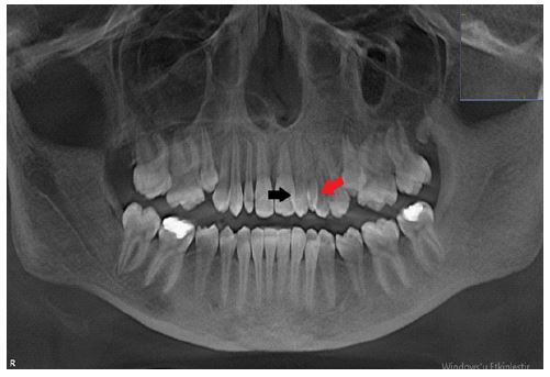
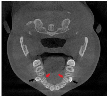
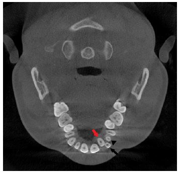
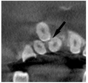
Table 1: Distribution and rates of cases with transposition in terms of jaw, classification in literature.
n %
Jaw
Maxilla 36 92.3
Mandible 3 7.7
Type
A-Mx.C.P1 17 47.6
B-Mx.C.I2 12 38.1
C-Mx.C to M1 0 0
D-Mx.I2.I1 1 2.4
E-Mx.C to I1 2 4.7
F-Mn.I2.C 3 7.2

Type A (Mx.C. P1): Upper canine-first premolar transposition.
Type B (Mx.C.12): Upper canine -lateral incisor transposition.
Type C (Mx.C to M1): Transposition of the upper canine to the first molar region.
Type D (Mx.12.11): Upper lateral incisor-central incisor transposition.
Type F (Mn.12.C): Lower lateral incisor-canine transposition.

Discussion

The prevalence of transpositions is reported to be approximately 0.4% in the literature [2,3]. In our study, the frequency of transposition was found to be 0.78%, higher than previous studies. This is the first study in which dental transpositions were examined in Turkish society by using CBCT.

In their study [9], reported that women were more frequently affected than men. In a study by [2], both genders were found to be affected equally (11 males and 10 females). In our study, the number of women who were found to have transposition was higher than the number of men (21 were female and 18 were male).

In [2] study, the range of age was found as 9-45 years, mean age was 17.7 years, mean age of women was 15.5 and the mean age of men was 19.7. In our study, transposition was found between the ages of 14 and 70. In parallel with the previously conducted studies, mean age of patients was 21.10; mean age of women was 23.47 and mean age of men was found as 18.33.

In the study by [9], the number of unilateral cases was much higher than the number of bilateral cases (61 unilateral, 8 bilateral cases). Similar to these studies, most of the dental transposition was unilateral (n=36, 92.3%).

Dental transpositions are mostly seen in the maxilla [10]. Shapira and Kuftinec [5] reported that 82% of transposed teeth were in the maxilla, while [10] reported that 83% of the cases were in the upper jaw. Our study was also in parallel with the literature, with 39 of the 36 transposed teeth were in the maxilla (92.3%).

Did [9] not find a significant difference in right-left distribution in unilateral cases (29 right and 32 left). Reported [2] that the cases in the right and left sides were equal in number, most of the maxillary transpositions were on the left side and all of the mandibular transpositions were on the right side. In our study, of the unilateral cases in maxilla, 20 were on the right, while 13 were on the left and all of the cases in the mandible were on the right side, in parallel with [2] study.

There are two types of transposition as complete or partial [5]. In [2] study, 15 of 22 cases were complete, 7 were partial transpositions. Found [11] the number of complete and partial transposition cases as equal. There were 16 complete and 26 partial transpositions in our study. It is thought that the reason for the difference in the number of complete and partial transpositions from the literature is the fact that panoramic radiography was used in the aforementioned studies, while evaluations were made with CBCT, which allowed for a more detailed examination, in our study.

In the transposition classification they conducted, [10] reported that most common type of transposition was between maxillary canine-premolar teeth (Type A-Mx.C.P1) (71%) [10,12], while [11]. Reported the most common type of transposition as maxillary canine- lateral dental transposition (Type B-Mx.C.12) (57.89%). In our study, similar to [10], the most common type of transposition was between maxillary canine-premolar transposition (Type A-Mx.C.P1) with 47.6% [10] reported the frequency of other transpositions as Type B (Mx.C.12) 20%, Type C (Mx.C to M1) 4%, Type D (Mx.12.11) 3%, Type E (Mx.C to 11) 2%. In our study, the order of frequency was as Type B (Mx.C.12) 38.1%; Type D (Mx.12.11) 2.4%; Type E (Mx.C to 11) 4.7%; Type F (Mn.12.C) 7.2%.

Transpositions not including canine tooth such as central-lateral incisory transpositions Type D (Mx.12.11) [6] and transpositions of maxillary canine and first molar (Type C-Mx.C to M1) are quite rare [13]. The least common transposition in this study was Type D and it was found only in one case. However, transposition between maxillary canine and first molar was not found in our study.

It has been reported that transpositions can be seen with dental anomalies such as wedge-shaped laterals (microdontia), congenital missing tooth, impacted teeth, persistent primary teeth, supernumerary teeth [2,9]. While [14] did not find a relationship between dental transposition and other anomalies, Shapira and Kuftinec [5] reported in their study that in 55.5% of the patients, dental transposition was seen together with one or more dental anomaly. In our study, similar to this study, dental anomalies other than transposition were found in the relevant side in 30 patients (76.92%). The relationship between dental transposition and persistent primary teeth has been described in literature, and primary teeth have been found to be persistent in transpositions related with permanent canines more frequently [15]. In our study, 27 persistent primary teeth were found in the relevant area in 24 patients (61.53%). Of the persistent primary teeth, 25 were primary canine, while 1 was primary lateral and 1 was primary second molar teeth.

In the study of [11], it was stated that the most frequently observed dental anomaly in all tooth transpositions was persistent primary teeth, and it was stated that 57.9% of 19 cases had persistent primary teeth. In our study, microdontia was found in 5.12%, impacted tooth was found in 5.12%, supernumerary teeth were found in 2.56%, 4 congenital missing teeth were found in 2 patients (two maxillary lateral and two maxillary second premolar teeth).

No data were found in literature on external root resorption in dental transposition cases. External root resorption was found in 7(17.94%) cases in our study. Pathologies such as external root resorption that can occur due to transposed teeth can only be examined with detailed tomographic images.

Conclusion

The prevalence of transposition was found as 0.78% in Turkish population. The present study is the first one to examine dental transpositions by using CBCT in Turkish society. External root resorption was found in some of the transposition cases. Routinely used two-dimensional imaging methods are not sufficient in the diagnosis of problems such as external root resorption that may be due to transposed teeth. For this reason, CBCT is essential in planning the treatment of transposition cases.

Declarations

Conflict of interest: Authors declare that there is no conflict of interest.

‘All procedures followed were in accordance with the ethical standards of the responsible committee on human experimentation (institutional and national) and with the Helsinki Declaration of 1975, as revised in 2008. Informed consent was obtained from all patients for being included in the study.’

Ethics committee approval: Ethics committee approval was not received since sources obtained from humans or animals were not used in this study.

Financial support: No financial support was received from any institution or organization for this study.

Consent to participate: Not Applicable.

Consent for publication: Not Applicable.

Availability of data and material: Data and materials are available.

Authors’ contributions: Kubra Cam wrote the main manuscript text; Ayse Zeynep Zengin performed the data collection and text editing. All the authors reviewed the manuscript.

The datasets generated and/or analyzed during the current study are available in the Ondokuz Mayıs University repository. The datasets used and/or analyzed during the current study are available from the corresponding author upon reasonable request.

References

  1. Peck L, Peck S, Attia Y. Maxillary canine-first premolar transposition, associated dental anomalies and genetic basis, Angle Orthod. 1993; 63: 99-109.
  2. Yılmaz HH, Türkkahraman H, Sayın M. Prevalence of tooth transpositions and associated dental anomalies in a Turkish population. Dentomaxillofacial Radiology. 2005; 5: 32-35
  3. Chattopadhyay A, Srinivas K. Transposition of teeth and genetic etiology. Angle Orthod. 1996; 66: 147-52.
  4. White SC, Pharoah MJ. Oral Radyoloji İlkeler ve Yorumlama. 7. Baskı. Ankara: Palme Yayınevi. 2018; 587-588.
  5. Shapira Y, Kuftinec MM. Maxillary tooth transpositions: Characteristic features and accompanying dental anomalies. Am J Orthod Dentofacial Orthop. 2001; 119: 127-34.
  6. Peck S, Peck L. Classification of maxillary tooth transpositions. Am J Orthod Dentofacial Orthop 1995; 107: 505-17.
  7. Joshi MR, Bhatt NA. Canine transposition. Oral Surg Oral Med Oral Pathol. 1971; 31: 49-54.
  8. Nambiar S, Mogra S, Shetty S. Transposition of teeth: A forensic perspective. Journal of Forensic Dental Sciences. 2014; 6: 151-153.
  9. Cho SY, Chu V, Ki Y. A retrospective study on 69 cases of maxillary tooth transposition. J Oral Sci. 2012; 54(2): 197-203.
  10. Peck S, Peck L, Kataja M. Mandibular lateral incisor-canine transposition, concomitant dental anomalies, and genetic control. Angle Orthod. 1998; 68: 455-66.
  11. Celikoglu M, Miloglu O, Oztek O. Investigation of tooth transposition in a non-syndromic Turkish anatolian population: Characteristic features and associated dental anomalies. Med Oral Patol Oral Cir Bucal. 2010; 15(5): e716-20.
  12. Danielsen JC, Karimian K, Ciarlantini R, Melsen B, Kjaer I. Unilateral and bilateral dental transpositions in the maxilla-dental and skeletal findings in 63 individuals. Eur Arch Paediatr Dent. 2015; 16: 467-76.
  13. Kayıpmaz S, Sarıcaoğlu ST, Sezgin ÖS. Unusual teeth transpositions: Two case reports, Oral Radiology. 2009; 25: 81-84.
  14. Papadopoulos MA, Chatzoudi M, Karagiannis V. Assessment of characteristic features and dental anomalies accompanying tooth transposition: A meta-analysis. Am J Orthod Dentofacial Orthop. 2009; 136(3): 308 e1-10; discussion 08-9.
  15. Bourzgui F, Sebbar M, Ait Ikiss J, et al. Tooth transposition: A descriptive study in a 547-patient sample. Int Orthod. 2012; 10(3): 311-7.