
Journal of Clinical Images and Medical Case Reports
ISSN 2766-7820
Case Series - Open Access, Volume 5
Case reports of bosonic pair annihilation on graphene by NaCl + KCl solution
*Corresponding Author : Chur Chin
Department of Emergency Medicine, New life Hospital, Bokhyun-dong, Bukgu, Daegu, Korea.
Email: nuestrodios@nate.com
Received : Oct 09, 2024
Accepted : Oct 25, 2024
Published : Nov 01, 2024
Archived : www.jcimcr.org
Copyright : © Chin C (2024).
Abstract
The peak at the binding energy of about 285 eV corresponds to the sp2 carbon atoms in graphene monolayer. About 100 eV is needed to make a pair production of boson particles. Therefore, graphene exfoliator, NaCl + KCl solution can use to make a pair annihilation of boson particles. Fermion needs lesser Fermi energy to make a pair production or a pair annihilation by dopped colloid gold. Colloid gold with camostat mesilate powder can disappear the tumor mass by the pair annihilation and inhibiting the chraomatin remodelling. Combination of NaCl + KCl solution and colloid gold with camostat mesilate powder can induce the quantum entanglement to re-appear the disease.
Citation: Chin C. Case reports of bosonic pair annihilation on graphene by NaCl + KCl solution. J Clin Images Med Case Rep. 2024; 5(11): 3322.
Introduction
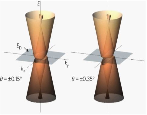
The electronic structure of epitaxial graphene is two dimensional by nature. It is a zero-gap semiconductor, i.e., a semimetal, with a conically shaped valence and conduction band reminiscent of relativistic Dirac cones for massless particles [1]. The hole doping in the conical band structure of epitaxial graphene monolayers can be achieved by the adsorption of gold. In the case of this doping, the Dirac point is shifted into the unoccupied states (Figure 2).
Quantum superposition is a fundamental principle of quantum mechanics that states that linear combinations of solutions to the Schrödinger equation are also solutions of the Schrödinger equation. This follows from the fact that the Schrödinger equation is a linear differential equation in time and position. The theory of quantum mechanics postulates that a wave equation completely determines the state of a quantum system at all times. If current tumor grading and staging system can be converted into this superposition concept,we can choose the treatment modalties and predict the prognosis more precisely [2].
Literature review
This paper is a review of the literature on theranostic effect of graphene based NaCl + KCl solution on cancer patients. We conducted a complete review of the available English literature, charting all reported cases and examining the treatment to outcome data.
Case reviews
Case 1: A 73-year-old elderly female patient presented with progressive dysphagia for more than 1 month with an Eastern Cooperative Oncology Group (ECOG) score of 1. After completing gastroscopy + pathological biopsy, chest enhanced CT, barium esophageal meal, PET-CT, and other related examinations, the clinical diagnosis was thoracic segmental esophageal poorly differentiated squamous carcinoma cT2N2M0 stage III. On February 16, 2023, and March 9, 2023, and 500 mL normal saline with 5cc KCl solution was injected for 6 hours intravenously. After the injection, the patient underwent an imaging examination. The chest enhanced CT suggested that the lesion range was significantly reduced compared with the previous scan, and mediastinal lymph nodes were partially reduced [3].
Case 2: A 34-year-old male patient with no significant medical history presented to a tertiary medical institution with generalized seizures. After management with anti-seizure medication, a neurological examination revealed no additional focal deficits. Brain MRI performed at the initial hospital showed an approximately 8 cm diffuse, infiltrative, non-enhancing mass lesion in the left temporal lobe. Based on clinical presentation and imaging features, a tentative diagnosis of low-grade glioma was made. Operation was done. The final pathology results revealed an isocitrate dehydrogenase (IDH)-mutant, CNS WHO grade 2 astrocytoma according to the 2021 classification system. 1 year after surgery, a brain MRI revealed an approximately 2 cm round, solid, enhancing mass in the left insular region, with perilesional T2 high signal intensity. Homogenous diffusion restriction with a mild increase in cerebral blood volume suggested tumor recurrence with high-grade transformation. The second operation was done. The final pathology results revealed an IDH-mutant, CNS WHO grade 4 astrocytoma according to the 2021 classification system. 4 days after surgery, he developed sudden right-side weakness and numbness, and right homonymous hemianopsia.Radiation therapy was not recommended and normal saline with KCl solution was injected. The patient recovered from the hemiparesis completely [4].
Case 3: A 27-year-old male patient, with a known history of Hodgkin lymphoma (HL), presented with gait disturbance. A brief summary of his HL history is as follows. Three years ago, he presented with persistent high fever and weight loss. Imaging revealed systemic lymphadenopathies in the axillary, neck, and mediastinal regions, as well as multifocal osseous lesions. A diagnostic biopsy from a cervical lymph node, coupled with a bone marrow assessment, confirmed a diagnosis of classic HL, lymphocyte-rich variant, at stage IV XB with documented bone marrow involvement. He received the NaCl + KCl solution intravenously, which successfully led to the absence of disease in the bone marrow and achieving a metabolic complete response [5].
Case 4: A 50-year-old adult patient with multiple brain and lung metastases (initially diagnosed as FIGO2018 stage IIA1) reported cough 2 years after postoperative chemoradiotherapy in Dec, 2022. Computed tomography (CT) of the thorax revealed multiple spaces occupying both lungs. The largest one was in the right lower lobe and measured 4 × 6 cm. A needle biopsy revealed squamous cell carcinoma with P16+ which supported the diagnosis of metastasis from multiple brain and lung metastases. No genetic, family, or psychosocial history were reported.She received the NaCl + KCl solution intravenously, After the injection, Magnetic resonance imaging (MRI) of the brain revealed a single, well-circumscribed peripheral-enhancing lesion measuring 3.6 × 2.5 cm within the left supratentorial lobe with surrounding vasogenic edema in Feb 2023 [6].
Case 5: A aged 34 with no particular pathological history, presented for one year with diabetes insipidus associated with a decrease in visual acuity evolving in a context of conservation of general condition. Cerebral MRI revealed a nodular lesion in the pineal region measuring 12.2 x 10.8 x 9.3 mm, associated with thickening of the pituitary stem suggestive of a germinoma. The patient underwent the NaCl + KCl solution intravenously, with a complete response on followup imaging [7].
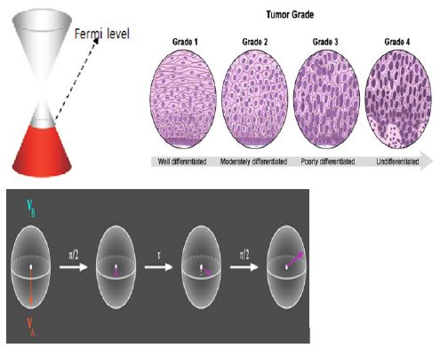
Superposition of tumor grading:
Grade1: 1-5 times of atypical cell is found from 20 biopsy samples.
Grade2: 5-10 times atypical cell is found from 20 biopsy samples.
Grade3: 10-15 times atypical cell is found from 20 biopsy samples.
Grade4: 15-20 times atypical cell is found from 20 biopsy samples.
Discussion
Superposition is the ability of a quantum system to be in multiple states at the same time until it is measured. The Bardeen–Cooper–Schrieffer (BCS) ground state is a superposition of different numbers of Cooper-paired electrons, not a superposition of different numbers of total electrons. The unpaired electrons are still there in the material, just not paired. To be consistent with spin statistics theorem, the wave function of the system must be antisymmetrized for fermions and symmetrized for bosons [8]. As a result of this symmetrization and antisymmetrization, it is indeed true that the expected value of the distance between particles is increased for fermions and reduced for bosons. The particles are indistinguishable, not only in their intrinsec properties, but are also sufficiently close for their wave function to overlap.
Remained massive or massless fermion superposition [9], which was not removed by the bosonic pair annihilation is seen on follow Computed Tomogram (CT) and Magnetic Resonance Image (MRI) or Positron Emission Tomogram (PET) scan. These remnants can be disappeared by colloid gold with camotstat powder intake 1 month after. The fermion remnant waves are disappeared by colloid gold with camotstat powder.
Conclusion
Graphene exfoliator, NaCl + KCl solution induce the bosonic pair annihilation as symmetrized state and colloid gold with camostat powder make the fermionic pair annihilation as asymmetrized state. Therefore, even after the bosonic pair annihilation by NaCl + KCl solution, the massless fermionic superposition can be remained. This fermionic remnant can be removed by colloid gold with camostat powder 1 month after.
References
- Isabella Gierz, Christian Riedl, Ulrich Starke, Christian R. Ast, and Klaus Kern. Atomic Hole Doping of Graphene, Nano Lett., 2008; 8(12): 4603-4607. DOI: 10.1021/nl802996s
- Pieter Kok WJ, Munro, Kae Nemoto. TC Ralph, Jonathan P. Dowling, and G. J. Milburn. Linear optical quantum computing with photonic qubits, Rev. Mod. Phys. 2007; 79: 135-174. DOI:https://doi.org/10.1103/RevModPhys.79.135
- Xiong Liu, Maoqi Wang, Deyuan Meng, Yuntao Tang , Qingtong Shi. Case report: A case study of neoadjuvant immunochemotherapy for locally advanced esophageal squamous carcinoma, Front Oncol. 2024; 14: 1332314. doi:10.3389/fonc.2024.1332314
- Byungjun Woo, Nayoung Han, Jeong Hoon Kim, Ho-Shin Gwa. Early High-Grade Transformation of IDH-Mutant Central Nervous System WHO Grade 2 Astrocytoma: A Case Report, Brain Tumor Research and Treatment 2024; 12(3): 186-191. DOI: https://doi.org/10.14791/btrt.2024.0022
- Hwanhee Lee, Sangjun Ahn, Seung Heon Cha, Won Ho Cho. Intracranial Involvement of Systemic Hodgkin Lymphoma: A Case Report and Literature Review, Brain Tumor Res Treat. 2024; 12(1): 63–69. doi: 10.14791/btrt.2023.0041
- Juan Ni, Xiaoyue DongXiaoyue Dong, Huafeng Shou, Qing XuQing Xu, Zhuomin Yin, Hanmei Lou. Second-line monotherapy with a PD-1/CTLA-4 inhibitor effectively treated multiple brain and lung metastases of cervical cancer: a case report, Front. Immunol. 2024; 15: 1-6. https://doi.org/10.3389/fimmu.2024.1434697
- Iharti R, Hadraoui G, Darfaoui M, Berkich S, Oumghar N, Elomrani A, et al. Double Localization of a Cerebral Germinoma: Case Report, Scholars Journal of Medical Case Reports. 2024; 12(5): 800-802. DOI: 10.36347/sjmcr.2024.v12i05.058
- Shufen Wang, Chao Wang and Xiang Ji. Towards understanding the salt-intercalation exfoliation of graphite into graphene, RSC advances. RSC Adv. 2017; 7: 522527-52260. DOI: 10.1039/c7ra07489a
- Keun Su Kim, Andrew L. Walter, Luca Moreschini, Thomas Seyller, Karsten Horn, Eli Rotenberg & Aaron Bostwick. Coexisting massive and massless Dirac fermions in symmetry-broken bilayer graphene, Nature Materials. 2013; 12: 887-892.