
Journal of Clinical Images and Medical Case Reports
ISSN 2766-7820
Clinical Image - Open Access, Volume 5
Worst nightmare of chronic silicosis
Beatriz Riquito1*; Mariana Certal1; Elisabete Cerqueira1; Sara Raimundo2; Victor Paz1
1Internal Medicine Unit, Chaves Hospital, Unidade Local de Saúde de Trás-os-Montes e Alto Douro, Portugal.
2Pneumology Unit, Vila Real Hospital, Unidade Local de Saúde de Trás-os-Montes e Alto Douro, Portugal.
*Corresponding Author : Beatriz Riquitoa
Internal Medicine Unit, Chaves Hospital, Unidade Local de Saúde de Trás-os-Montes e Alto Douro, Portugal.
Tel: +351961482777;
Email: beatrizriquito@gmail.com
Received : Oct 15, 2024
Accepted : Oct 31, 2024
Published : Nov 07, 2024
Archived : www.jcimcr.org
Copyright : © Riquito B (2024).
Abstract
Simultaneous bilateral pneumothorax is an uncommon presentation and usually occurs in the context of secondary spontaneous pneumothorax. A 45-year-old man diagnosed with complicated silicosis for, with a history of numerous pneumothorax requiring inpatient treatment, is readmitted to the hospital with a new left pneumothorax. After 7 day with a pleural drain, a chest radiography showed a right pneumothorax. A computed tomography confirmed a bilateral pneumothorax, a rare complication of silicoses, requiring thoracic surgery to definitive management.
Keywords: Silicosis; Bilateral pneumothorax; Pleural drain; Thoracic surgery.
Citation: Riquito B, Certal M, Cerqueira E, Raimundo S, Paz V. Worst nightmare of chronic silicosis. J Clin Images Med Case Rep. 2024; 5(11): 3331.
Introduction
Silicosis is one of the most frequent occupational diseases caused by inhalation of small particules of crystalline silica. Simultaneous bilateral pneumothorax is an uncommon presentation and usually occurs in the context of secondary spontaneous pneumothorax. The association of pneumothorax and silicosis is infrequent and most cases are unilateral. Bilateral pneumothorax in patients with silicosis is very rare with just a few reports in medical literature.
Case presentation
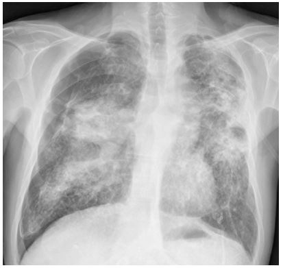
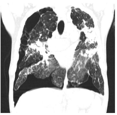
This case presents a 45-year-old man diagnosed with complicated silicosis for, with a history of numerous pneumothorax requiring inpatient treatment, either left or right-sided, always managed with pleural drains and pleurodesis. He is readmitted with a new left pneumothorax, initially managed with a pleural drain. Seven days later, a control X-ray showed not only the left pneumothorax not yet resolved, but also a right pneumothorax (Figure 1), confirmed later with computed tomography (Figure 2). After a second pleural drain was inserted, the patient was later referred to a specialized center where he was later submitted to thoracic surgery as a definitive treatment.
Conclusion
Secondary spontaneous pneumothorax are associated with chronic silicosis with massive advanced fibrosis and, in most cases, occur unilaterally. Bilateral pneumothorax is rarely seen and mainly observed on accelerated silicosis. It responds very poorly to non-surgical therapy (pleural drains and/or pleurodesis), leaving video-assisted-thoracoscopic surgery (VATS) as the only viable option.
References
- Meena MK, Singh R, Joshi N, Rathore SS, Chadalawada S, et al. Silicosis with Secondary Spontaneous Pneumothorax in the Western Rajasthan. Cureus. 2020; 12(11): e11811.
- Fotedaar S, Chaudhary D, Singhla V, Narang R. Silicosis with bilateral spontaneous pneumothorax. Lung India. 2010; 27(3): 173-175.
- Siburian RN, Dewi KP, Koesoemoprodjo W. Diagnostic approach and management of bilateral pneumothorax due to silicosis in Indonesian male: A rare case. Int J Surg Case Rep. 2022; 97: 107407.