
Journal of Clinical Images and Medical Case Reports
ISSN 2766-7820
Clinical Image - Open Access, Volume 5
A young lady with painful fingers and abnormal fundus
Nisahan B1*; Dayaratna J2; Peranantharajah T3
1Senior Registrar, Department of Medicine, Teaching Hospital Jaffna, Jaffna, Sri Lanka.
2Registrar, Department of Medicine, Teaching Hospital Jaffna, Jaffna, Sri Lanka.
3Consultant Physician, Department of Medicine, Teaching Hospital Jaffna, Jaffna, Sri Lanka.
*Corresponding Author : Nisahan B
Senior Registrar, Department of Medicine, Teaching Hospital Jaffna, Jaffna, Sri Lanka.
Email: nisahan2004@yahoo.com
Received : Oct 28, 2024
Accepted : Nov 14, 2024
Published : Nov 21, 2024
Archived : www.jcimcr.org
Copyright : © Nisahan B (2024).
Citation: Nisahan B, Dayaratna J, Peranantharajah T. A young lady with painful fingers and abnormal fundus. J Clin Images Med Case Rep. 2024; 5(11): 3351.
Description
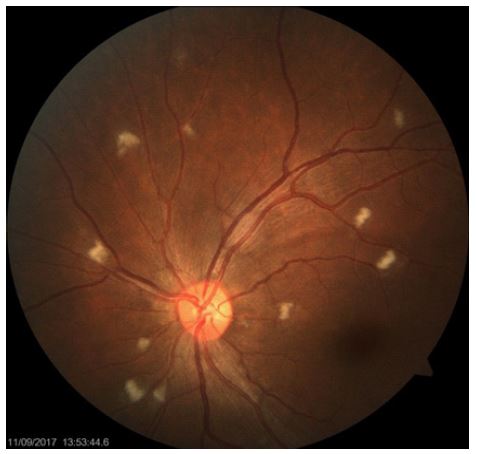
A 36 years old previously healthy lady presented with painful 2nd and 5th fingers of both hands for 6 months duration. The pain had gradually got worse and was found to have blackish complexion of the fingers suggestive of gangrene. There were no joint involvements or any skin changes. There was significant hair loss during last few months. Her urine output was normal and there is mild facial swelling and bilateral ankle oedema. There were no abnormalities in respiratory or cardiovascular system. Ophthalmoscopic examination revealed multiple cotton wool spots (Figure 1).
Investigations showed, bicytopenia with microcytic anemia of HB% 7.8 g/L and platelets count of 95000/μL. Inflammatory markers were elevated with ESR 110 mm in 1st hour, CRP 45 mg/dl. Renal function tests were normal. Urine full analysis revealed proteinuria, without any active urinary sediment. Liver tests showed low albumin of 15 g/L and normal range transaminases. Ultrasound scan of abdomen was normal and renal biopsy revealed histological diagnosis of WHO class 1V lupus nephritis. ANA and DsDNA antibodies were positive but cANCA and pANCA were negative. Diagnosis of Systemic Lupus Erythematosus was confirmed and patient was managed with intravenous steroids, intravenous cyclophosphamide and hydroxychloroquine.
Although SLE can affect any part of the eye, keratoconjunctivitis sicca being the most common manifestation as a result of secondary Sjögren’s disease [1]. The next most common pathologic condition involving the eye in lupuѕ patients is retinal vasculopathy in the form of cotton wool spots. Cotton-wool spots represent edematous and ischemic neuronal tissue [2]. Systemic hypertension and diabetes mellitus are the commonest causes for cotton wool spots in the fundus. In addition it can occur due to ischemic, embolic, infectious, toxic, pancreatitis, immune-mediated, radiation-induced, neoplastic, traumatic, tractional and idiopathic causes [3]. For clinicians with limited resources, early detection of this condition provides confidence in the clinical diagnosis, resulting in improved outcomes from early care.
References
- Silpa-Archa S, Lee JJ, Foster CS. Ocular manifestations in systemic lupus erythematosus. British Journal of Ophthalmology. 2016; 100(1): 135-41.
- Lanham JG, Barrie T, Kohner EM, Hughes GR. SLE retinopathy: Evaluation by fluorescein angiography. Annals of the Rheumatic Diseases. 1982; 41(5): 473-8.
- Arroyo JG. Cotton-wool spots may challenge diagnosis. Rev Ophthalmol. 2004; 11: 4.