
Journal of Clinical Images and Medical Case Reports
ISSN 2766-7820
Short Report - Open Access, Volume 5
Galeazzi equivalent fracture in children: A case report
Hind Abou El Jaoud1,2*; Tayeb Bentayeb1,2; Lamiae Chater1,2
1Department of Pediatric Surgery, University Hospital Mohamed VI, Tangier, Morocco.
2Faculty of Medicine and Pharmacy of Tangier, Abdelmalek Essâadi University, Tangier, Morocco.
*Corresponding Author : Hind Abou El Jaoud
Department of Pediatric Surgery, University Hospital Mohamed VI, Tangier, Morocco.
Tel: +21-2670500457;
Email: h.aboueljaoud@uae.ac.ma
Received : Oct 22, 2024
Accepted : Nov 15, 2024
Published : Nov 22, 2024
Archived : www.jcimcr.org
Copyright : © El Jaoud HA (2024).
Citation: El Jaoud HA, Bentayeb T, Chater L. Galeazzi equivalent fracture in children: A case report. J Clin Images Med Case Rep. 2024; 5(11): 3353.
Description
Fractures of the forearm are frequently observed in children and adolescents. Among these, the Galeazzi fracture-characterized by a fracture of the radial shaft accompanied by Dislocation of the Distal Radioulnar Joint (DRUJ)-is relatively uncommon in this age group. The Galeazzi equivalent fracture is a variation that presents in pediatric populations as a radius fracture associated with a displaced physeal injury of the distal ulna, but without DRUJ dislocation.
We report a case of a 15-year-old patient with no significant surgical history who sustained an injury while playing football. The patient presented with right wrist pain. Physical examination revealed swelling of the wrist with no skin lacerations and a limited range of motion. The neurovascular assessment was unremarkable.
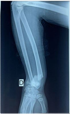
Radiographic evaluation (posterior-anterior view) of the right wrist demonstrated a transverse no displaced fracture of the distal third of the radius and a distal ulnar fracture classified as Salter-Harris type IV. Following a trial of closed reduction, subsequent X-rays indicated a displaced and unstable fracture pattern.
The radial shaft fracture was successfully reduced and stabilized, but the ulnar fracture remained unstable, necessitating fixation with a 1.5 mm smooth K-wire. Post-fixation X-rays confirmed proper alignment of both fractures.
The patient was monitored postoperatively at two, four, and six weeks. At the three-week follow-up, the splint was replaced with a below-elbow cast. The K-wires were removed after six weeks. Complete bone union was achieved, with the patient demonstrating a normal range of motion six months postoperatively.
Conclusion
In conclusion, anatomic reduction is crucial for patients with malalignement, especially older patients with lower potential for adequate bone remodeling. Regular follow-up is essential to monitor for growth arrest and other potential complications.