Journal of Clinical Images and Medical Case Reports
ISSN 2766-7820
Clinical Image - Open Access, Volume 5
Lung herniation - A rare cause of chest pain
Diana Amorim*; Sónia Silva
Pulmonology Service, Local Health Unit of Leiria, Rua das Olhalvas, Pousos, 2410-197 Leiria, Portugal.
*Corresponding Author : Amorim D
Pulmonology Service, Local Health Unit of Leiria, Rua das Olhalvas, Pousos, 2410-197 Leiria, Portugal.
Email: dianasofiaamorim@gmail.com
Received : Nov 03, 2024
Accepted : Nov 19, 2024
Published : Nov 26, 2024
Archived : www.jcimcr.org
Copyright : © Amorim D (2024).
Citation: Amorim D, Silva S. Lung herniation-A rare cause of chest pain. J Clin Images Med Case Rep. 2024; 5(11): 3358.
Description
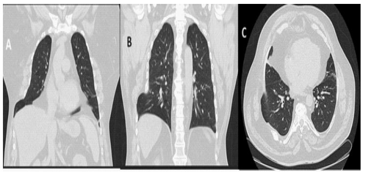
Lung hernias are a rare entity that results from the protrusion of part of the lung parenchyma through the rib cage. It can occur after trauma or as a post-surgical iatrogenic, but is even less common when it occurs spontaneously. Clinically, the most common symptom is chest pain, sometimes with the presence of a hematoma at the site, so clinicians should be aware of these signs and symptoms, even though they are nonspecific. The authors present a clinical case of a 57-year-old man, a former smoker, who was overweight and had obstructive sleep apnea. He came to the Emergency Room (ER) with one-week history of dry cough, with no other associated symptoms, no alterations on physical examination or significant analytical changes. Viral infection was assumed and the patient was discharged home. He returned five days later with chest pain in the right latero-basal region, worsened by coughing, and the presence of chest wall swelling in the same location. Objective examination revealed the presence of a hematoma at that region and no other significant alterations. A chest CT scan was requested, which showed a fracture of the 7th costal arch and exuberant right lung herniation in the same area (Figure 1). Given that there was no history of trauma or previous surgery, this herniation was assumed to be due to coughing.
The patient was then followed up in a pulmonology consultation. He maintained chest pain and a hematoma in the chest wall, with difficulty in activities of daily living and was afterward referred for thoracic surgery. Although surgical treatment is being considered less and less, it should be an option in the event of significant symptoms, with excellent results and low morbidity. This case highlights the importance of an extremely rare entity that can become disabling.