
Journal of Clinical Images and Medical Case Reports
ISSN 2766-7820
Case Report - Open Access, Volume 5
Diabetic ketoacidosis with intra-abdominal hernia: A rare cause of small bowel perforation - A case report
Shixiao Dong1#; Mengrui Chai2,3,4,5#; Wenshan Liu2,3,4,5; Xin Zhang2,3,4,5; Fuqiang Liu2,3,4,5*
1Department of Emergency, Qilu Hospital of Shandong University, Jinan, Shandong 250012, People’s Republic of China.
2Department of Endocrinology and Metabolism, Qilu Hospital of Shandong University, Jinan, Shandong 250012, People’s Republic of China.
3Shandong Provincial Key Laboratory of Spatiotemporal Regulation and Precision Intervention in Endocrine and Metabolic Diseases, Jinan, Shandong 250012, People’s Republic of China.
4Shandong Provincial Engineering Research Center for Advanced Technologies in Prevention and Treatment of Chromic Metabolic Diseases, Jinan, Shandong 250012, People’s Republic of China.
5Institute of Endocrine and Metabolic Diseases of Shandong University, Jinan, Shandong 250012, People’s Republic of China.
#Shixiao Dong and Mengrui Chai contributed equally to this work.
*Corresponding Author : Fuqiang Liu
Department of Endocrinology and Metabolism, Qilu Hospital of Shandong University, Jinan, Shandong 250012, People’s Republic of China.
Email: liufuqiang@sdu.edu.cn
Received : Nov 03, 2021
Accepted : Nov 21, 2024
Published : Nov 28, 2024
Archived : www.jcimcr.org
Copyright : © Fuqiang L (2024).
Abstract
Background: Diabetic Ketoacidosis (DKA) is a lifethreatening medical condition. Intra-abdominal hernia causes intestinal obstruction that results in a severe acute abdomen; if it is not diagnosed and treated in time, it can result in serious complications such as intestinal strangulation necrosis, intestinal perforation, and even septic shock, resulting in death. Especially in patients with DKA, intra-abdominal hernia is rare and lacks specific clinical symptoms and auxiliary examination methods; therefore, early diagnosis is important to reduce mortality and morbidity.
Case presentation: Here, we present a case of a 51-year-old Chinese male with diabetes for more than 10 years who was admitted to our hospital due to DKA. DKA caused a rare intra-abdominal hernia, which resulted in intestinal perforation and intra-abdominal infection. Emergency surgery was performed to remove part of the small intestine and abdominal wall of the small intestine. After active rescue treatment, the patient was discharged in stable condition.
Conclusion: In patients with DKA, intra-abdominal hernia can result in serious complications such as intestinal perforation and intra-abdominal infection, and therefore, early diagnosis and treatments are required to reduce mortality and morbidity.
Keywords: Diabetic ketoacidosis; Intra-abdominal hernia; Intestinal rupture; Intra-abdominal infection; Case report.
Abbreviations: DKA: Diabetic Ketoacidosis; DM: Diabetes Mellitus; CT: Computed Tomography; VAC: Vacuum Assisted Closure; ICU: Intensive Care Unit; ABAH: Adhesion Band Abdominal Hernia.
Citation: Shixiao D, Mengrui C, Wenshan L, Xin Z, Fuqiang L. Diabetic ketoacidosis with intra-abdominal hernia: A rare cause of small bowel perforation - A case report. J Clin Images Med Case Rep. 2024; 5(11): 3362
Background
An intra-abdominal hernia is defined as the dislocation of the internal abdominal organs from their original position through a normal or abnormal orifice or fissure in the abdominal cavity to an abnormal space. It is a rare cause of intestinal obstruction [1]. Diabetes Mellitus (DM) is a serious disease threatening human health; however, patients’ awareness of the disease is still low, and some patients do not adhere to their treatment regimens and healthy lifestyle behaviors. Diabetic Ketoacidosis (DKA) is a common clinical acute complication of diabetes. Previous research found that the prevalence of DKA was high in patients with DM and COVID-19 or even new-onset DM following COVID-19; since the COVID-19 pandemic, DKA has attracted the attention of researchers [2]. DKA aggravates the original symptoms of DM, with non-specific clinical manifestations, such as abdominal pain. To ensure the health and safety of DKA patients with abdominal pain, relevant examinations should be performed to identify the cause of abdominal pain while actively treating ketoacidosis. Here, we discuss a patient with DKA and intra-abdominal hernia who underwent emergency surgery.
Case presentation
A 51-year-old Chinese male (height, 1.73 m; weight, 74 kg; and body mass index, 24.72 kg/m2) with diabetes for more than 10 years was admitted to the emergency department of our hospital due to lower abdominal pain, nausea, and vomiting for 5 days, and the patient developed dizziness and fatigue 1 day prior to admission. The patient was diagnosed with DKA based on the following laboratory results: glucose, 19.8 mmol/l; lactate, 4.9 mmol/l; anion gap, 32.6 mEq/l; and positive serum ketones. Then the patient was managed through nil per oral, adequate intravenous fluid supplement, anti-infection, correction of electrolyte imbalance, hemostasis, and nutritional support treatment. After his condition stabilized, the patient was transferred to the endocrine department of our hospital for further diagnosis and treatment.
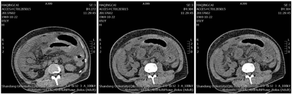
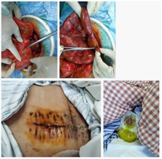
On the second day after admission, at 01:40 AM., the patient complained of pain in the upper abdomen and around the umbilical cord and discharged dark red watery stools, with colic and pale complexion; his blood pressure was 98/74 mmHg. At 02:03 AM., the patient vomited dark green fluid, with intermittent diarrhea, and his abdominal pain aggravated. The patient was given pain relief, acid suppression, and fluid rehydration treatment. Later, the patient’s condition continued to worsen, and his abdominal pain could not be relieved. Having no clear exacerbating factor for his DKA, in conjunction with persisting epigastric pain and nausea, abdominal-pelvic Computed Tomography (CT) was ordered. CT examination showed abdominal pelvic effusion and gas under the diaphragm and abdominal cavity; diagnostic peritoneal puncture showed dark brown ascites (Figure 1). Color Doppler ultrasound showed a large amount of fluid in the abdominal cavity. Based on the above presentation, gastrointestinal perforation, abdominal infection, and septic shock were diagnosed. After evaluation, oral tracheal intubation was performed, and laparotomy was performed under general anesthesia to release adhesion, partially resect the small bowel, and reverse small bowel abdominal wall atrophy. The closure of the laparotomy wound was performed using Vacuum-Assisted Closure (VAC) suction. The small intestine seen during the surgery is shown in (Figure 2). The patient was transferred to the intensive care unit after the surgery, where he was treated for 33 days (days 3-35 after admission). His blood pressure and heart rate decreased, and he had fever, bilateral pleural effusion and partial lung insufficiency, lung fungal infection, and gastrointestinal hemorrhage. A multidisciplinary team was consulted because small bowel incarceration would cause ischemia, necrosis, and perforation of this segment of the bowel, leading to complex abdominal infection, sepsis, and multi-organ failure. After symptomatic diagnosis and treatments, such as chest compressions, epinephrine, alkali supplementation, and anti-infection, the patient’s condition gradually stabilized. On the 36th day of admission, the patient was transferred back to the endocrinology ward to continue rehabilitation, where he was treated for 5 days. Then, he was discharged from the hospital on the 41st day.
Discussion
The causes of DKA in diabetic patients include infection, discontinuation of treatment, and stress. In diabetic patients, DKA can cause fatal complications if it is not diagnosed and treated promptly [3]. The main diagnostic criteria for DKA in modern medicine are as follows: ① blood glucose >11.0 mmol/L or history of diabetes; ② blood ketone body >3.0 mmol/L or urine ketone body above 2+; and ③ blood bicarbonate concentration < 15 mmol/L and/or venous blood pH < 7.3 [4]. The reported patient had diabetes, normal treatment was not regular, and blood sugar control was not satisfactory; the patient discontinued his treatment before the onset of DKA. Laboratory tests revealed that the pH value was reduced and that urine ketone bodies were positive. Based on these results, the patient was diagnosed with DKA. Relevant recommendations for the treatment of DKA suggest volume restoration with isotonic fluid, ensuring serum potassium >3.3 mmol/L before initiation of insulin therapy of 0.1 unit/kg body weight, while bicarbonate infusion should be considered in cases of life-threatening acidosis with pH <6.8 [5]. The cause of our DKA patient’s abdominal pain was unclear at the initial stage of hospitalization. The patient’s abdominal pain persisted even after the correction of ketoacidosis, which was accompanied by symptoms such as abdominal distension, nausea, and vomiting. The level of white blood cells had increased significantly, and D-dimer and fibrinogen levels had also increased. It is difficult to rely on DKA “monism” to explain the persistent abdominal pain; therefore, it is imperative to actively search for the cause of abdominal pain [6]. The causes of abdominal pain in patients with DKA may be as follows: ① High concentration of hydrogen ions stimulate the gastrointestinal mucosa nerve or destroy the gastrointestinal mucosa to cause inflammation; the lower the pH and carbon dioxide binding capacity in the serum, the higher the concentration of H﹢, thereby causing abdominal pain. ② Electrolyte disturbances such as low potassium and low sodium cause gastrointestinal cramps, gastric dilatation, and even paralytic intestinal obstruction, leading to abdominal pain. ③ Studies have shown that acute hyperglycemia can cause gastrointestinal disorders and gastrointestinal motility disorders and that gastric emptying is delayed; under stress conditions, intestinal endotoxin stimulation and Oddi sphincter contraction will cause increased pressure in the gallbladder and bile ducts, leading to abdominal pain. ④ the accumulation of metabolic toxic products leads to “pseudoperitonitis” [7]. In DKA patients, insulin is further deficient or impaired; peripheral glucose utilization decreases; fat mobilization is forced to increase; free fatty acids increase significantly, leading to hyperlipidemia; and a large number of acidic metabolites, such as acetoacetate, acetone, and β-hydroxybutane, are produced. These acidic metabolites stimulate the peritoneum and induce abdominal pain [8]. An intestinal/internal adhesion band abdominal hernia is defined as the formation of intra-abdominal adhesion frenulum due to various reasons and the formation of local fissures or cavities [9]. Factors such as strenuous activity and trauma increase the intra-abdominal pressure. The atypical secondary intra-abdominal hernia formed by the protrusion of the peristaltic intestines and other structures herniating into the space result in a rare clinical acute abdomen. Surgery is the only effective treatment method. The basis for effective treatment lies in the early and correct diagnosis of the disease. The clinical manifestations are non-specific, and the clinical knowledge is insufficient. Early accurate diagnosis before surgery is difficult, and the rate of misdiagnosis is high. Our patient had an adhesion band abdominal hernia. At the same time, DKA caused intestinal edema, dilation, and paralysis and finally caused intra-abdominal hernia, resulting in acute intestinal compression, necrosis, and rupture. Finally, emergency surgery was performed to remove the necrotic intestine and intestinal abdominal wall fistula, saving the patient’s life. The closure of the laparotomy wound was performed using VAC suction. The condition of DKA, which is mainly manifested by abdominal pain, is more serious than common DKA, but it is often missed and misdiagnosed during the examination [10]. Therefore, medical staff should pay particular attention to the identification of intestinal obstruction and intestinal rupture [11]. DKA abdominal pain is different from acute abdomen; in patients with DKA, the initial symptoms include thirst, urination, nausea, vomiting, sweating, chest tightness, and autonomic nervous disorders, followed by abdominal pain and deep and fast breathing, and their breath smells like rotten apples. Our patient initially had pain in the upper abdomen and around the umbilical cord; after 23 minutes, he began to have nausea and vomited dark green fluid, with intermittent diarrhea. The symptoms of the patient suggested surgical acute abdomen, such as intestinal obstruction, intra-abdominal hernia, and intestinal perforation. Then, a CT examination was performed in time to obtain a clear diagnosis. Once intra-abdominal hernia causes intestinal obstruction, it is difficult to restore the herniated intestinal segment through conservative treatment. The only effective treatment is to operate as soon as possible. Timely surgical treatment can reduce the incidence of intestinal necrosis and increase the success rate of treatment [12].
Conclusion
In short, for DKA patients with non-specific symptoms, such as abdominal pain, clinical diagnosis is difficult, and the rate of misdiagnosis is high. The occurrence of intestinal obstruction caused by intra-abdominal hernia in DKA patients is rare in clinical practice. The etiology is complicated, with non-specific clinical manifestations, and the preoperative diagnosis is difficult. Multi-slice spiral CT can be used as the preferred imaging method to improve the preoperative diagnosis rate of the disease. Early diagnosis and timely surgery are key to successful treatment, which can avoid strangulation and necrosis of the intestinal wall to a certain extent and improve the prognosis of patients.
Declarations
Author contributions: All authors have read and approved the final version of the manuscript.
Funding: No funding was received.
Availability of data and materials: The data generated in the present study may be requested from the corresponding author.
Conflict of interest: The authors declare that they have no conflict of interest.
Patient consent for publication: Written informed consen was obtained from the patient for publication of this case report and any accompanying images.
Ethical approval: Ethical approval for the study was granted by the Qilu Hospital of Shandong University Ethics Committee. All procedures performed in studies involving human participants were in accordance with the ethical standards of the institutional and/or national research committee and with the 1964 Helsinki Declaration and its later amendments or comparable ethical standards.
References
- Hola J, Azolas R, Abedrapo M, Avillo V, Sobrón M, et al Internal hernia secondary to a broad ligament defect Rev Chil Cirug. 2010; 62: 13-4.
- Joustra ML, Raidt JJ, Droog F, Veneman TF. Diabetic Ketoacidosis, Hypertriglyceridemia and Abdominal Pain due to Acute Pancreatitis Complicated by Non-immune Haemolytic Anaemia. European journal of case reports in internal medicine. 2020; 7(12): 002085.
- Mondal S, DasGupta R, Lodh M, Gorai R, Choudhury B, et al. Predictors of new-onset diabetic ketoacidosis in patients with moderate to severe COVID-19 receiving parenteral glucocorticoids: a prospective single-centre study among Indian type 2 diabetes patients. Diabetes Metab Syndr. 2021; 15(3): 795-801.
- Chinese Society of Diabetes. Guidelines for the prevention and treatment of type 2 diabetes mellitus in China (2020 edition) [J]. Chinese Journal of Diabetes Mellitus. 2021; 13(4): 315.
- Sampani E, Sarafidis P, Dimitriadis C, Kasimatis E, Daikidou D, et al. Severe euglycemic diabetic ketoacidosis of multifactorial etiology in a type 2 diabetic patient treated with empagliflozin: Case report and literature review. BMC Nephrol. 2020; 21: 276.
- Margekar S, Jayant S, Jatav O, Jain M, Chouksey A. Abdominal pain in diabetes--DKA is not the only cause. The Journal of the Association of Physicians of India. 2014; 62(5): 450-451.
- Karamanos E, Dulchavsky S, Beale E, Inaba K, Demetriades D. Diabetes Mellitus in Patients Presenting with Adhesive Small Bowel Obstruction: Delaying Surgical Intervention Results in Worse Outcomes. World journal of surgery. 2016; 40(4): 863-869.
- Lax Y, Singh A. Referred Abdominal Pain. Pediatrics in review. 2020; 41(8): 430-433.
- Nakamura T, Sato T, Naito M, Ogura N, Yamanashi T, et al. Laparoscopic Surgery is Useful for Preventing Recurrence of Small Bowel Obstruction After Surgery for Postoperative Small Bowel Obstruction. Surg Laparo Endo Per. 2016; 26(1): 1-4.
- Fields JM, Dean AJ. Systemic causes of abdominal pain. Emergency medicine clinics of North America. 2011; 29(2): 195-210.
- Urru A, Romano N, Melani EF, Rollandi GA. An unusual cause of large bowel perforation: ingestion of a clam valve. Internal and Emergency Medicine. 2020;16(1): 225-226.
- Shek K, Ditkofsky N, Ashamalla S, Patel C. A case report of caecal herniation through the foramen of Winslow. Annals of the Royal College of Surgeons of England. 2018; 100(6): 142-144.