
Journal of Clinical Images and Medical Case Reports
ISSN 2766-7820
Case Report - Open Access, Volume 5
Prenatal ultrasound diagnosis of Dandy-Walker syndrome at 22 weeks of gestation: A case report
Subash Pandey; Sushmita Bhattarai*; Prasiddha Khatri
KIST Medical College and Teaching Hospital, Gwarko, Lalitpur, Nepal.
*Corresponding Author : Sushmita Bhattarai
KIST Medical College and Teaching Hospital, Gwarko, Lalitpur, Nepal.
Email: sushmita.bhattarai12@gmail.com
Received : Nov 02, 2024
Accepted : Nov 22, 2024
Published : Nov 29, 2024
Archived : www.jcimcr.org
Copyright : © Bhattarai S (2024).
Abstract
Dandy-Walker Malformation (DWM) is a rare congenital brain disorder characterized by cerebellar vermis hypoplasia, fourth ventricle enlargement, and cystic dilation in the posterior fossa. We report a 23-year-old primigravida at 22 weeks and 5 days gestation, presenting with persistent vomiting. Anomaly scan showed a cystic structure (15x9x9 mm) between the fourth ventricle and cisterna magna, with cerebellar vermis hypoplasia, indicating DWM. After counseling, medical induction was performed. The patient was discharged in stable condition, with contraceptive counseling, and declined karyotyping. This case highlights the value of prenatal diagnosis and counseling in DWM management.
Keywords: Dandy walker malformation; Hydrocephalus; Medical induction.
Abbreviations: DWM: Dandy Walker Malformation; CNS: Central Nervous System; USG: Ultrasonography; RBC: Red Blood Cells; WBC: White Blood Cells; PV: Per Vaginal; MRI: Magnetic Resonance Imaging; BPD: Bi-parietal Diameter; HC: Head Circumference.
Citation: Pandey S, Bhattarai S, Khatri P. Prenatal ultrasound diagnosis of Dandy-Walker syndrome at 22 weeks of gestation: A case report. J Clin Images Med Case Rep. 2024; 5(11): 3364.
Introduction
Dandy-Walker Malformation (DWM) is a rare congenital CNS disorder involving cerebellar vermis underdevelopment, fourth ventricle enlargement, and posterior fossa expansion. Caused by genetic and environmental factors, it has an incidence of 1 in 25,000-35,000. DWM is more common in females, with a male-to-female ratio of 1:3 reported in a Spanish study [1]. Dandy-Walker malformation’s causes are not fully understood, but genetic aberrations, syndromic malformations, and congenital infections are thought to contribute. It accounts for about 3% of hydrocephalus cases [2]. DWS is typically confirmed by MRI or postnatal pathology, but due to poor fetal prognosis, early diagnosis is advised [3]. In ultrasonography, DWM is marked by ventriculomegaly, choroid plexus cyst, meningocele, ventricular septal defect, abnormal heart development, small aortic arch,endocardial cushion absence, complex cardiovascular malformations, gallbladder enlargement, digestive tract obstruction, limb shortening, polydactyly, hydronephrosis, kidney enlargement, neck cyst, polyhydramnios, thickened nuchal translucency, umbilical hernia, and umbilical cord cyst [4]. Ultrasound accurately diagnoses DWM and should prompt screening for CNS, systemic anomalies, and chromosomal abnormalities, as prognosis depends on associated malformations and karyotype [2].
Case report
A 23-year-old primigravida with a last menstrual period on 2024-03-22 presented to the Gynecology OPD at 22 weeks and 5 days with persistent, intermittent vomiting for one month (1-2 episodes every other day). The vomiting was non-progressive, non-projectile, and not bile or blood-stained, with no abdominal pain or other symptoms. She was taking iron and calcium regularly and reported normal fetal movements. On examination, the patient was alert, oriented, and in fair condition. No pallor, icterus, edema, clubbing, cyanosis, dehydration, or palpable lymph nodes were found. Respiratory, cardiovascular, and CNS exams were normal. Abdominally, the uterus matched 20 weeks gestation, with palpable fetal parts and a fetal heart rate of 138 bpm.
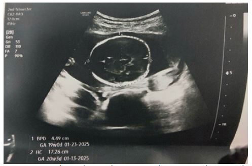
Table 1: USG findings.
| Anomaly scan | USG findings |
|---|---|
| Fetus Number | 1 |
| Presentation, Movements | Breech, Normal |
| Placental Site | Fundo-posterior |
| Placental Thickness | Normal |
| Cord Vessels Number | 3 |
| Liquor Amount | Normal |
| Heart | Heart Rate: 150 beats/minute, Heart enlarged with asymmetrical cardiac chambers |
| Diaphragm, stomach, Abdominal Wall, Kidneys, Bladder, Cranium | Normal |
| Bowel | Echogenic Bowel loops noted |
| Cerebellum | Hypoplastic cerebellar vermis |
| Ventricles | Normal |
| Spine | Normal |
| Fetal Upper Limbs | Short for gestational age |
| Fetal Lower Limbs | Short for gestational age |
Discussion
Dandy-Walker syndrome is a rare congenital disorder marked by cerebellar vermis underdevelopment, fourth ventricle enlargement, and an enlarged posterior fossa. Risk factors include infections (toxoplasmosis, CMV, rubella) and drug exposure (warfarin, isotretinoin, ethanol). In this case, the mother had no such exposures. Dandy-Walker Syndrome can be diagnosed early using high-resolution fetal ultrasound and MRI if needed [1-5]. In our case, prenatal diagnosis was done through Ultrasonographic analysis twice on 30th August and 19th september, 2024 [6]. Ultrasound shows cystic enlargement of the fourth ventricle, cerebellar vermis hypoplasia, and ventricular dilation in Dandy-Walker Malformation (DWM). MRI can offer further details. A USG on 19th September 2024 showed an 18-week 5-day fetus with a cystic structure (15x9x9 mm) connecting the fourth ventricle to the cisterna magna and vermis hypoplasia. DWM is often associated with congenital heart disease and neural tube defects [7]. Dual marker screening showed a short nasal bone (4.3 mm), depressed nasal bridge, and nuchal fold thickness of 3.6 mm, suggesting a congenital anomaly. The 30th August USG revealed an enlarged heart with asymmetrical chambers, echogenic bowel loops, and short fetal long bones. The 19th September USG showed mildly echogenic bowel loops and a 1.8 mm echogenic focus in the right ventricle. Limb abnormalities have occurred as the less common abnormalities. In our case the fetal long bones were short for gestational age [6-8]. This finding was helpful in prenatal diagnosis of DWS.
Conclusion
This case highlights the importance of early prenatal diagnosis of Dandy-Walker malformation for informed decision-making. Detailed fetal ultrasound aids in identifying associated anomalies, informing prognosis and management. Parental counseling is crucial for understanding DWM’s implications and guiding decisions. Comprehensive prenatal care, including genetic counseling, provides critical information for ethical, patient-centered clinical decisions.
Declarations
Consent: Written informed consent was obtained from the patient for publication and any accompanying images.
Competing interests: The authors declare no competing interests.
Source of funding: None.
References
- Hamid HA. Dandy-Walker Malformation.
- Abdul H, Burns J, Estevez A, Nasr El-Nimer C, Ekinde B, et al. Hemorrhagic Stroke in a Young Adult with Undiagnosed Asymptomatic Dandy–Walker Malformation. Case Rep Neurol Med. 2019; 1-3.
- Sun Y, Wang T, Zhang N, Zhang P, Li Y. Clinical features and genetic analysis of Dandy-Walker syndrome. BMC Pregnancy Childbirth. 2023; 23(1): 40.
- Shaffer LG, Rosenfeld JA, Dabell MP, Coppinger J, Bandholz AM, et al. Detection rates of clinically significant genomic alterations by microarray analysis for specific anomalies detected by ultrasound. Prenat Diagn. 2012; 32(10): 986-95.
- Ndu I, Chinawa J, Chikani M, Ibekwe R, Aronu A, et al. Dandy Walker malformation (variant): late presentation with childhood blindness. Niger J Paediatr. 2014; 42(1): 73.
- Harper T, Fordham LA, Wolfe HM. The fetal dandy walker complex: associated anomalies, perinatal outcome and postnatal imaging. Fetal Diagn Ther. 2007; 22(4): 277-81.
- Murray JC, Johnson JA, Bird TD. Dandy-Walker malformation: etiologic heterogeneity and empiric recurrence risks. Clin Genet. 1985; 28(4): 272-83.
- Salihu HM, Kornosky JL, Druschel CM. Dandy-Walker syndrome, associated anomalies and survival through infancy: a population-based study. Fetal Diagn Ther. 2008; 24(2): 155-60.