Journal of Clinical Images and Medical Case Reports
ISSN 2766-7820
Clinical Image - Open Access, Volume 5
A case of neonatal necrotizing enterocolitis
Li Shenmei*; Cai Xianhu; Huang Dong; Peng Yiwen; Qiu Yucheng
Radiology Department, Meizhou Maternal and Child Health Hospital, Meizhou, Guangdong, China.
*Corresponding Author : Li Shenmei
Radiology Department, Meizhou Maternal and
Child Health Hospital, Meizhou, Guangdong, China.
Email: 2551614908@qq.com
Received : Nov 21, 2024
Accepted : Dec 06, 2024
Published : Dec 13, 2024
Archived : www.jcimcr.org
Copyright : © Shenmei L (2024).
Abstract
Neonatal Necrotizing Enterocolitis (NEC) is a common disease in newborns with a relatively high clinical incidence. Premature infants with NEC are mainly associated with prematurity, low birth weight, immature development, and non-breastfeeding. The incidence of NEC in full-term infants is low, accounting for about 5% to 25% of all NEC cases, and its mechanism of occurrence is different from that of premature infants, mainly related to birth asphyxia, congenital heart disease, hypoglycemia, sepsis, etc.
Keywords: NEC; Neonatal; X-ray.
Citation: Shenmei L, Xianhu C, Dong H, Yiwen P, Yucheng Q. A case of neonatal necrotizing enterocolitis. J Clin Images Med Case Rep. 2024; 5(12): 3386.
Introduction
Early or mild NEC typically presents with non-specific findings on abdominal X-rays, which may sometimes only manifest as changes in bowel motility, intestinal distension, bowel obstruction, the degree of bowel rigidity and dilation, and blurring and widening of the bowel spaces. Subsequently, as the disease progresses, bright shadows such as linear, strip-like, beaded, and arc-shaped can be seen in the intestinal wall on X-ray plain films. Portal venous gas appears as bright shadows extending from the hepatic hilum outward, resembling the shape of dead branches.
Case description
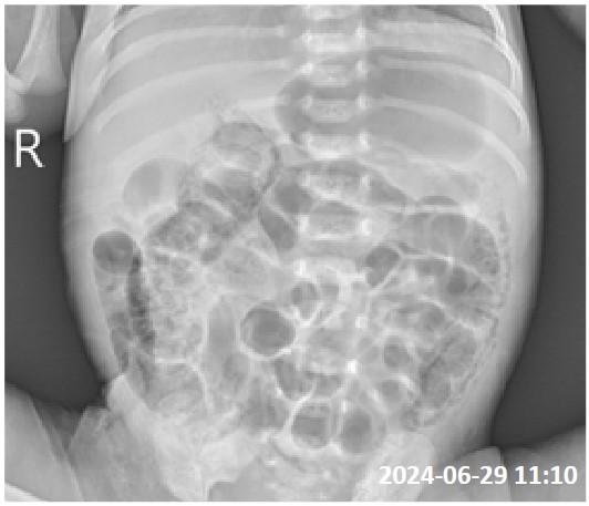
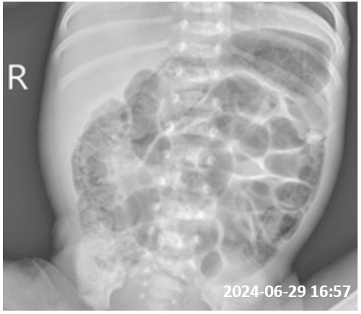
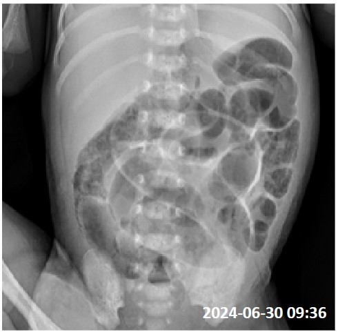
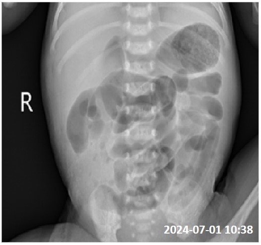
The premature baby was born at our hospital on 2024-06-25 at 35+1 weeks of gestation, with poor life ability. On the evening of 2024-06-29, the baby had poor feeding and vomited stomach contents once. The bowel sounds were weak. Abdominal X-ray plain films were taken on and 2024-07-01.
Comment
NEC Phase I- The bedside X-ray of the patient shows only intestinal dilation and gas accumulation or slightly irregular intestinal morphology, and mild intestinal obstruction can also be seen. NEC Phase II- The bedside X-ray of the patient may show irregular shape of the intestinal tract, widened intestinal space, and thickened intestinal wall. More than two ographs suggest a relatively fixed course of the intestinal tract. It can present with intestinal wall gas accumulation, portal vein gas accumulation, and ascites. NEC Phase III- In addition to the above manifestations, a significantly increased density in the abdomen and blurred disappearance of the abdominal fat line indicating the presence of ascites, perforation signs are often present, and free gas in the abdominal cavity can be seen on X-ray.