
Journal of Clinical Images and Medical Case Reports
ISSN 2766-7820
Case Report - Open Access, Volume 2
Chest CT surprising improvement in COVID-19 patient
Elisa Calabrò1*; Claudia Gabiati1; Gabriella Nucera2; Valentina Raffaelli2; Francesco Bombaci3; Antonio Brucato1
1Internal Medicine, ASST Fatebenefratelli Sacco, Milano, Department of Biomedical and Clinical Sciences, University of Milan, Italy.
2 UOC Medicina D’Urgenza, ASST Fatebenefratelli Sacco, Milano, Italy.
3 Radiology Department, ASST Fatebenefratelli Sacco, Milano, Italy.
*Corresponding Author : Elisa Calabrò
Internal Medicine, Ospedale Fatebenefratelli e Oftalmico, ASST Fatebenefratelli-Sacco, Piazza Principessa
Clotilde 3, 20121 Milano, Italy.
Email: elisa.calabro@asst-fbf-sacco.it
Received : Apr 07, 2021
Accepted : Apr 23, 2021
Published : Apr 27, 2021
Archived : www.jcimcr.org
Copyright : © Calabrò E (2021).
Abstract
A surprising improvement in COVID-19 chest CT follow up gives hope regarding future clinical evolution of COVID-19 patients.
Citation: Calabrò E, Gabiati C, Nucera G, Raffaelli V, Bombaci F, et al. Chest CT surprising improvement in COVID-19 patient. J Clin Images Med Case Rep. 2021; 2(2): 1071.
Case report
A 56-year-old man was admitted for severe COVID-19 pneumonia [1-3]. He did not smoke and was on treatment with valsartan 80 mg daily for hypertension. Body mass index was 32.7. Symptoms (Cough and Fever) started 8 days before hospital admission. Nasopharyngeal RT-PCR swab was positive for SARS CoV-2. Laboratory results showed increased white blood cells count, C-Reactive Protein (CRP), lactate dehydrogenase, D-dimer and fibrinogen values (11.6 x 10⁹ /L, normal range 4.19- 9.35; 79 mg/L, normal range 0-5; 251 U/L, normal range 135-225; 1794 ng/ml; 600 mg/dl normal range 270-470; respectively). He was treated with hydroxychloroquine, lopinavir/ritonavir and steroids association. Ten days after admission he experienced a rapidly worsening of respiratory exchanges (P/F ratio 40) and needed endotracheal intubation. Contrast enhanced Chest CT scan was immediately done and pulmonary embolism was excluded. Selective right broncus intubation was noted (Figure 1 panel C), and the tube was retracted accordingly. Axial views at apexes, aortic arch, carina bifurcation, main right scissura and up the diaphragm, showed an almost complete bilateral pulmonary consolidation, with poorly represented ground glass opacities (Figure 1 panels A,B,C,D,E). Bronchial aspirate culture done the same day was negative, and no clear evidence of secondary infection was present; broad spectrum antibiotics were started. CRP the day after intubation was 53 mg/L.
After 4 days in the Intensive Care Unit, clinical condition progressively improved and ventilatory support was gradually weaned.
A CT scan done 16 days after the previous one showed a surprising improvement, with impressive reduction of opacities and consolidations; some ground glass opacities and reticular pattern were still visible, with predominant peripheral and posterior location (Figure 1 panels F,G,H,I,L). CRP was 6 mg/L.
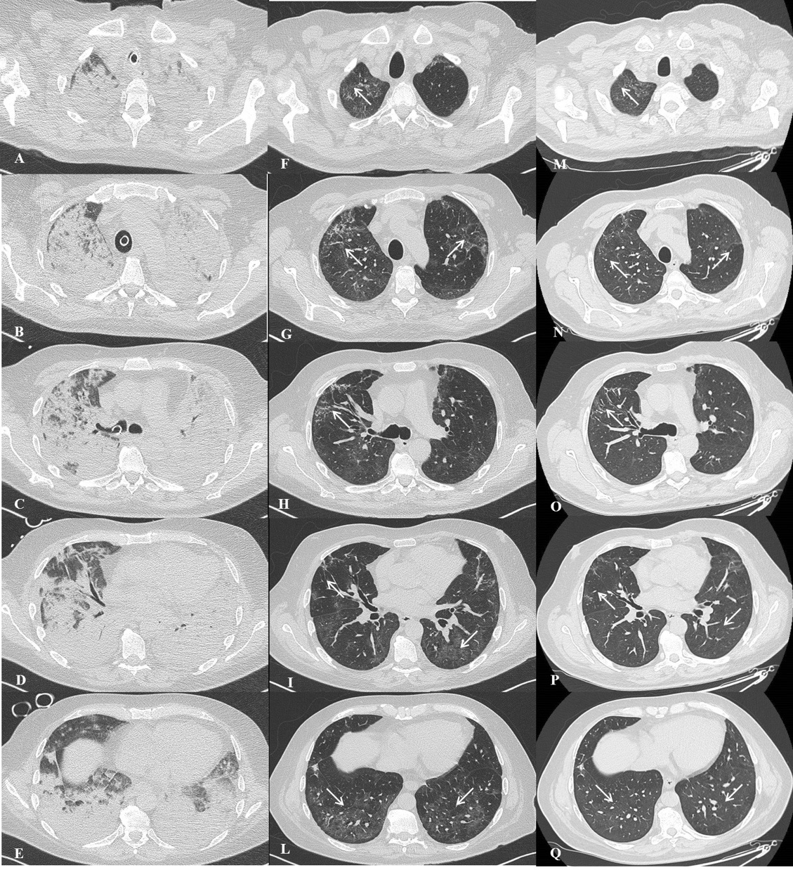
Thirty-nine days after admission, patient was transferred in a rehabilitation unit, clinically cured and with a P/F ratio of 276. Fifty day after the second CT (67 days after admission) he performed a follow-up chest CT that showed markedly reduction in ground glass opacities and reticular lesions previously described (Figure 1 panels M,N,O,P,Q). Post-discharge lung function evaluation, performed 67 days after admission, demonstrated a mild restriction and reduction in diffusing capacity of the lung for carbon monoxide (TLC 78% predicted and DLCO 71% predicted).
Conclusion
The striking radiological and clinical improvement gives hope regarding future clinical evolution of this and other subjects.
Contributorship statement: Elisa Calabrò, Claudia Gabiati, Gabriella Nucera, Valentina Raffaelli, Antonio Brucato have actively participated in the preparation of the article and have read and approved the text. Francesco Bombaci performed supplemental chest CT and radiological revision.
Funding: The authors have not declared a specific grant for this research from any funding agency in the public, commercial or not-for-profit sectors.
References
- Carlos WG, Dela Cruz CS, Cao B, Pasnick S, Jamil S. Novel Wuhan (2019-nCoV) Coronavirus. Am J Respir Crit Care Med. 2020; 201: P7-8.
- Shi H, Han X, Jiang N, Cao Y, Alwalid O, Gu J et al. Radiological findings from 81 patients with COVID-19 pneumonia in Wuhan, China: A descriptive study. Lancet Infect Dis. 2020.
- Chung M, Bernheim A, Mei X, Zhang N, Huang M, Zeng X et al. CT Imaging Features of 2019 Novel Coronavirus (2019-nCoV). Radiology. 2020; 295: 202-207.