
Journal of Clinical Images and Medical Case Reports
ISSN 2766-7820
Case Report - Open Access, Volume 2
A case report of periapical cemento-osseous dysplasia
Murude Akyolal
Department of Oral and Maxillofacial Surgery, Biruni University, Istanbul, Turkey
*Corresponding Author : Eduardo dos Santos Sousa
Department of Oral and Maxillofacial Surgery, Biruni
University, Istanbul, Turkey
Email: murudeyazan@hotmail.com
Received : Mar 24, 2021
Accepted : Apr 26, 2021
Published : Apr 29, 2021
Archived : www.jcimcr.org
Copyright : © Akyolal M (2021).
Abstract
Periapical Cemento-Osseous Dysplasia (PCOD) is one of the classified lesions of cemento-osseous dysplasia that generally consisted in middle aged black women. This article reported a case of a 36-yearold woman who was incidentally recognized with PCOD on the basis of panoramic radiograph.
According to the clinical and radiographical findings of the patient, diagnosis of PCOD was done. Treatment was not considered. Regular radiographical follow-up was recommended to the patient.
Keywords: Cemento-osseous dysplasia; Panoramic; Non-odontogenic tumor.
Citation: Akyolal M. A Case Report of Periapical Cemento-osseous Dysplasia. J Clin Images Med Case Rep. 2021; 2(2): 1081.
Introduction
Cemento-Osseous Dysplasia (COD) is the most common fibro-osseous lesion encountered in clinical practice and can be seen at the tooth bearing or edentulous areas of the jaws [1]. COD is represented as a benign lesion arising from undifferentiated cells of the periodontal ligament tissues[1]. Different reactive, dysplastic and neoplastic cells are seen by replacing with normal bone cells microscopically with a collagen matrix containing the trabeculae of immature bone and, cement-like hard tissue at fibro-osseous lesions [2].
COD is sub-divided into Periapical Cemento-Osseous Dysplasia (PCOD), focal cemento-osseous dysplasia and florid cemento-osseous dysplasia [3]. PCOD characteristically includes the apices of the teeth, typically one or more mandibular incisors [4].
PCOD occurs more frequently in women of black race older than 30 years of age [5,6].
This lesion is randomly seen in radiographs taken for other reasons. PCOD lesions are mostly have well-defined borders [7].
Typically, osteolytic; cementoblastic; and mature stages develop serially at this lesion [8]. Radiological appearance of the lesion depends on the stage of observation. In the osteolytic stage, a circular radiolucent lesion occurs at the apex of the root; in the second or cementoblastic stage, spicules of cement are defined because of cementoblastic activity increase, so that mixed appearence is observed radiographically. A completely radiopaque lesion appears at the final or mature stage [9-11].
Dentists should be aware about these incidentally defined lesions with appropriate radiological and clinical examination. Generally, no treatment is required and only regular followup is advised [12]. This report is about a case of PCOD that consulted to our hospital for dental problems which were not related with that lesion. Dental treatments of the patient were finished and regular followup is advised.
Case report
A 36-year-old woman was referred to the oral surgery clinic of Oral Dental Health Hospital in Kucukcekmece, Istanbul for mandibular third molar pain.
She reported that she didn’t have any systemic diseases. She had no history of trauma to the mandible. She didn’t have any symptoms such as pain, swelling in the extraoral examination. Left mandibular third molar was painful and had deep caries in the intraoral examination. But the other teeth and oral mucosa were healthy. A radiolucent-radiopaque mixed lesion was seen at mandibular anterior region in the radiological examination (Figure 1). Lesion was located on the roots of lower incisors and canines. Root resorption or tooth displacement didn’t seen at related teeth.
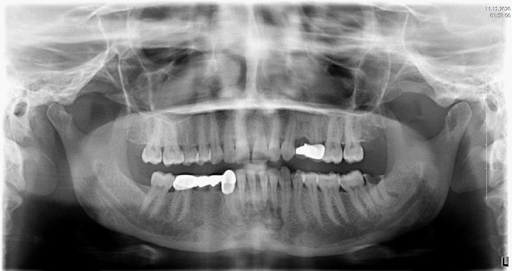
The lamina dura of involved teeth was dissapeared. Expansion of periodontal ligament space was found.
Periodontal tissues of mandibular anterior teeth were healthy. The teeth that related with lesion were vital in an electric stimulation test and they were asymptomatic.
According to the clinical and radiographical findings of the patient, diagnosis of PCOD was done. Treatment was not considered. Regular radiographical follow-up was recommended to the patient.
Discussion
COD is the most common fibro-osseous lesion of the jaws that strongly specified at middle-aged women of African descent [13].
The classification of COD remains unclear. Recently, the fourth edition of the World Health Organization Classification of Head and Neck Tumors transformed to the terminology “Cemento- Osseous Dysplasia” from “Osseous Dysplasia” for defining these tumors as odontogenic, with their origin of periodontal ligament [3].
COD is a group of fibro-osseous lesions that is sub-divided into Periapical Semento-Osseous Dysplasia (PCOD), focal semento-osseous dysplasia, and florid semento-osseous dysplasia.
PCOD characteristically includes the roots of the one or more mandibular incisors. Focal COD is a single lesion generally not related with a tooth, and fluoride COD includes two or more quadrants of the jaws [3,4].
COD includes of gritty tan-brown parenchyma histologically. Sub-divided three forms are histologically similar [14]. Pathological formation mechanism of these lesions is reactive and dysplastic changes that characterized by the replacement of microscopically normal bone with a collagen matrix including immature bone trabeculae and cement-like hard tissue [15]. Some groups defined that COD originates from the cells of the periodontal ligament [13].
These lesions don’t have any symptoms generally and incidentally identified on radiographs. Teeth related with PCOD area are generally vital in an electric stimulation test. Conversely, teeth having periapical infection are not viable. Fluoride COD can expand and therefore present with pain, especially if the lesion becomes infected. When Fluoride COD enlarges and becomes infected, pain cystic changes may present because of the lesion [16-18].
Genetic predisposition has not been defined for COD so far. Rarely familial forms with autosomal dominant transference have been reported named as “Familial Florid Osseous Dysplasia” or “Familial gigantiform sementoma [17,18]. These forms are commonly seen in the upper jaw and conversely other semento-osseous dysplasias that are more common in whites.
PCOD is a benign lesion that known to processed from undifferentiated cells of the periodontal ligament tissue pathologically [5]. PCOD is a common form of COD that mostly seen at the anterior mandible related with single or multiple teeth. Periapical COD occurs more frequently in women of black race [6].
Asymptomatic lesions can be single or multiple and do not cause changes in periodontal tissue. The lesions are widespread in the anterior region of the mandible near the apex of the mandibular incisors and canines, and the included teeth remain vital [2,8].
The lesions typically evolves in three stages: Osteolytic; sementoblastic; and mature [10]. Radiological image is attached to the moment of phase. In the first or osteolytic phase, an unilocular radiolucent lesion may be seen at the apex of the root; In the second or cementoblastic stage, spicules occur because of the sementoblastic activity, so the radiolucent lesion gets a mixed appearance [9,11]. The last or mature phase leads to a completely radiopaque lesion. Bone marrow spaces get more dense and sclerotic in late stage of disease.
The lesions typically evolves in three stages: Osteolytic; sementoblastic; and mature [10]. Radiological image is attached to the moment of phase. In the first or osteolytic phase, an unilocular radiolucent lesion may be seen at the apex of the root; In the second or cementoblastic stage, spicules occur because of the sementoblastic activity, so the radiolucent lesion gets a mixed appearance [9,11]. The last or mature phase leads to a completely radiopaque lesion. Bone marrow spaces get more dense and sclerotic in late stage of disease.
Calcified lesion can reach up to 10 cm diameter and is usually encircled by a radiolucent halo [19]. This development can take months or years, and during its development the diameter of the lesion increases from 0.2 cm to 10 cm or more [6].
Commonly, no treatment is required and only regular followup examinations are advised [12]. Biopsy of cemento-osseous dysplasia, particularly large and sclerotic lesions can lead to infection and secondary osteomyelitis of the mandible that can be difficult to treat with antibiotics because the bone is sclerotic and relatively avascular [20].
This report describes a case of periapical COD and discusses differential diagnosis. The teeth remain vital throughout the evolution of the lesion to the sclerotic stage and should not be extracted or treated endodontically. So, we didn’t treated this patient and we advised regular follow-up examinations.
Dentists should be aware about these asemptomatic lesions at routine radiological examinations.
References
- Eversole R, Su L, El Mofty SK. Benign fibro-osseous lesions of the craniofacial complex. A review. Head Neck Pathol. 2008; 2: 177- 202.
- Kawai T, Hiranuma H, Kishino M, Jikko A, Sakuda M. Cementoosseous dysplasia of the jaws in 54 Japanese patients: A radiographic study. Oral Surg Oral Med Oral Pathol Oral Radiol Endod. 1999; 87: 107-114.
- Mofty SK, Nelson B, Toyosawa S. Fibro-osseous and osteochondromatous lesions. In: WHO Classification of Head and Neck Tumors. Lyon: IARC Press. 2017: 251–255.
- Das BK, Das SN, Gupta A, Nayak S. Florid cemento-osseous dysplasia. J Oral Maxillofac Pathol. 2013; 17: 150.
- Manganaro AM and Millett GV. Periapical cemental dysplasia. Gen Dent1996; 44: 336-339.
- Eskandarloo A and Yousefi F: CBCT findings of periapical cemento-osseous dysplasia: A case report. Imaging Sci Dent. 2013; 43: 215-218.
- Alsufyani NA, Lam EW. Cemento-osseous dysplasia of the jaw bones: key radiographic features. Dentomaxillofac Radiol. 2011; 40: 141-146.
- Komabayashi T and Zhu Q: Cemento-osseous dysplasia in an elderly Asian male: a case report. J Oral Sci. 2011; 53: 117-120.
- Summerlin DJ and Tomich CE: Focal cemento-osseous dysplasia: A clinicopathologic study of 221 cases. Oral Surg Oral Med Oral Pathol. 1994; 78: 611-620.
- Scholl RJ, Kellett HM, Neumann DP and Lurie AG: Radiographics. Cysts and cystic lesions of the mandible: clinical and radiologichistopathologic review. Radiographics. 1999; 19: 1107-1124.
- Ariji Y, Ariji E, Higuchi Y, Kubo S, Nakayama E and Kanda S: Florid cemento-osseous dysplasia. Radiographic study with special emphasis on computed tomography. Oral Surg Oral Med Oral Pathol. 1994; 78: 391-396.
DiFiore P, Bowen S. Cemento-osseous dysplasia in AfricanAmerican men: A report of two clinical cases. J Tenn Dent Assoc. 2010; 90: 26-29.
Fenerty S, Shaw W, Verma R, Syed AB, Kuklani R, Yang J, Ali S. Florid cemento-osseous dysplasia: review of an uncommon fibro-osseous lesion of the jaw with important clinical implications. Skeletal Radiol. 2017; 46: 581–590.
Sedano HO, Kuba R, Gorlin RJ. Autosomal dominant cemental dysplasia. Oral Surg Oral Med Oral Pathol. 1982; 54: 642–646.
Coleman H, Altini M, Kieser J, Nissenbaum M. Familial florid cemento-osseous dysplasia–A case report and review of the literature. J Dent Assoc S Afr. 1996; 51: 766–770.
Mahomed F, Altini M, Meer S, Coleman H. Cemento-osseous dysplasia with associated simple bone cysts. J Oral Maxillofac Surg. 2005; 63: 1549–1554.
Sedano HO, Kuba R, Gorlin RJ. Autosomal dominant cemental dysplasia. Oral Surg Oral Med Oral Pathol. 1982; 54: 642–646.
Coleman H, Altini M, Kieser J, Nissenbaum M. Familial florid cemento-osseous dysplasia–a case report and review of the literature. J Dent Assoc S Afr. 1996; 51: 766–770.
MacDonald-Jankowski DS: Florid cemento-osseous dysplasia: A systematic review. Dentomaxillofac Radiol. 2003; 32: 141-149.
Regezi J, Sciubba J, Jordan R. Benign non-odontogenic tumors. In: Regezi J, Sciubba J, Jordan R, editors. Oral pathology: Clinical pathology correlates. 6th ed. Amsterdam: Elsevier. 2012; 294– 298.