
Journal of Clinical Images and Medical Case Reports
ISSN 2766-7820
Case Report - Open Access, Volume 2
Persistent iris vessels in a case of aniridia
Rashmi Deshmukh; Madhavan Rajan*
Department of Ophthalmology, Addenbrookes Hospital, Cambridge University Hospitals, Cambridge, United Kingdom.
*Corresponding Author : Madhavan Rajan
Department of Ophthalmology, Addenbrookes
Hospital, Cambridge University Hospitals, CambridgeCB20QQ, United Kingdom.
Email: madhavan.rajan@addenbrookes.nhs.uk
Received : Mar 27, 2021
Accepted : Apr 23, 2021
Published : Apr 26, 2021
Archived : www.jcimcr.org
Copyright : © Rajan M (2021).
Keywords: Aniridia; Persistent iris vessels; Iris remnants.
Citation: Deshmukh R, Rajan M. Persistent iris vessels in a case of aniridia. J Clin Images Med Case Rep. 2021; 2(2): 1083.
Description
Congenital aniridia is caused by a mutation in the PAX6 gene [1] and is characterized by partial or complete absence of iris tissue. Apart from the hypoplasia of iris tissue, other ocular features such as foveal hypoplasia, nystagmus, aniridia-related keratopathy, Peters anomaly, Axenfeld-Rieger anomaly and glaucoma are seen in these eyes [1,2]. Cases have been reported with persistent pupillary membranes [3] and iris strands [4].
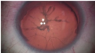
We report a case of a 27-year old female with aniridia where persistent iris vessels were noted. The vascular plexus was not visualized on slit lamp examination, however, when the patient was undergoing cataract surgery, the fragile capillaries were visualized under the operating microscope (Figure). There are few reports [3,5] of free-floating iris vessels visualized in the anterior chamber of patients with aniridia. Interestingly, similar to our case, Theobald et al [5] also noted the vascular plexus under the operating microscope. The persistent iris vessels were not visualized in the initial examination.
It is important to be mindful of finding these persistent iris vessels in cases with aniridia as these capillaries are at risk of rupture and causing hyphema [5]. Although the pathophysiology remains unknown, it has been suggested that the presence of this arcade of blood vessels originating from the small iris remnants at the angle, points towards a neuroectodermal defect causing aniridia [3].
References
- Lim HT, Kim DH, Kim H. PAX6 aniridia syndrome: clinics, genetics, and therapeutics. Curr Opin Ophthalmol. 2017; 28: 436–447.
- Pujari R, Deshmukh R, Rajan MS. Maternal finger-prick allogenic blood for persistent corneal epithelial defects. BMJ Case Reports.
- Hamming N, Wilensky J. Persistent pupillary membrane associated with aniridia. Am J Ophthalmol. 1978; 86: 118–120.
- Beauchamp GR, Meisler DM. An alternative hypothesis for iris maldevelopment (aniridia). J Pediatr Ophthalmol Strabismus. 1986; 23: 281–283.
- Theobald T, Davitt BV, Shields SR. Hemorrhagic glaucoma in an infant with hemophilia, spontaneous hyphema, aniridia, and persistent iris vessels. J AAPOS. 2001; 5: 129–130.