
Journal of Clinical Images and Medical Case Reports
ISSN 2766-7820
Case Report - Open Access, Volume 2
Synchronous HPV positive bilateral palatine tonsillar squamous cell carcinoma – Brief review of current literature on the role of magnetic resonance imaging
Mudit Arora1*; M Reza Taheri2; Arjun Joshi3; Stephanie Barak4
1 Fellow, Department of Radiology, George Washington University and Hospital, Washington DC, USA.
2 Associate Professor, Department of Radiology, George Washington University and Hospital, Washington DC, USA.
3 Professor, Department of Otolaryngology, George Washington University and Hospital, Washington DC, USA.
4 Assistant Professor, Department of pathology, George Washington University and Hospital, Washington DC, USA.
*Corresponding Author : Mudit Arora
Department of Radiology, George Washington University and Hospital, Washington DC, USA.
Email: drmuditarora@gmail.com
Received : Apr 05, 2021
Accepted : Apr 30, 2021
Published : May 05, 2021
Archived : www.jcimcr.org
Copyright : © Arora M (2021).
Abstract
Synchronous bilateral palatine tonsillar Squamous Cell Carcinoma (SCC) is a rare presentation and less than 10 cases of Human Papillomavirus (HPV) mediated synchronous bilateral palatine tonsillar SCC has been reported. We report case of a patient who presents with a painless left sided neck swelling and was subsequently diagnosed by imaging and histopathology with bilateral Squamous Cell Carcinoma (SCC) of the palatine tonsil and cervical nodal metastasis. Due to its excellent soft tissue/tumor contrast resolution, Magnetic Resonance Imaging (MRI) is the preferred imaging modality for initial staging. Improved sensitivity for the detection of synchronous tumors can be achieved by Positron Emission Tomography (PET)/ Computed Tomography (CT) examination. Due to the rare nature of this pathology, no specific treatment guideline has been provided by National Comprehensive Cancer Network (NCCN). Treatment options available include bilateral Transoral Robotic Surgery (TORS) radical tonsillectomy combined with bilateral neck dissection with adjuvant radiotherapy or chemoradiotherapy and/or upfront concurrent chemoradiotherapy.
Citation: Arora M, Taheri MR, Joshi A, Barak S. Synchronous HPV positive bilateral palatine tonsillar squamous cell carcinoma – Brief review of current literature on the role of magnetic resonance imaging. J Clin Images Med Case Rep. 2021; 2(3): 1109.
Background
Palatine tonsils are the most common site of oropharyngeal malignancy followed by the base of tongue and the soft palate [1]. The incidence of oropharyngeal malignancy associated with Human Papillomavirus Infection (HPV) has increased in the recent years, with approximately 40-80 percent of cases attributed to HPV [2,3].
Synchronous Head and Neck Squamous Cell Carcinoma (HNSCC) is defined as two foci of SCC, separated by a wide zone of uninvolved tissue, diagnosed concomitantly or within the first 6 months of each other. The term “Metachronous” is used for malignant tumors diagnosed between 6 months and 5 years after the diagnosis of the initial primary malignancy [3-5]. The estimated overall incidence of synchronous and metachronous HN-SCC malignancy is 1-8 percent and 17-30 percent, respectively. However, synchronous bilateral palatine tonsillar SCC is more unusual with less than 40 cases reported in the literature, with the first case reported in 1971 [6]. Moreover, less than 10 cases of HPV mediated synchronous bilateral palatine tonsillar SCC have been reported [7].
The role of imaging in palatine tonsil carcinoma is to radiologically stage the lesion based on its size and locoregional spread and to define the extent of the nodal spread. Although both contrast enhanced CT and MRI are routinely used for the staging of this cancer, MRI is the preferred modality due to its superior soft tissue/contrast resolution. PET-CT is a useful adjuvant, although tonsil tissue is normally FDG avid.
We present case of synchronous HPV mediated bilateral palatine tonsillar SCC and review the role of MR imaging in this diagnosis and its treatment.
Case report
A man in his early seventies presents with an insidious and painless left sided neck swelling. Patient reports no pain, dysphagia, odynophagia, hemoptysis, and denies any systemic complaints such as fever, fatigue, chills, nausea or weight loss. He has not noticed any new oral cavity ulcers/lesions.
His social history is remarkable for no history of smoking or prior tobacco exposure. He has no prior medical history and no prior history of radiation.
Investigations
On physical examination, enlarged nodes are palpable on the left side of his neck. Additionally, a heterogeneous mass is noted in the left palatine tonsil. The right palatine tonsil appears normal. A biopsy of the left palatine tonsil reveals HPV mediated p16+ fragments of squamous cell carcinoma, moderately to poorly differentiated, non-keratinizing in background of tonsillar tissue. The patient is counselled regarding the results and is scheduled for an MRI and a PET/CT exam. The patient is also advised to undergo a direct laryngoscopy under general anesthesia for further characterization and surgical planning.
MRI reveals a heterogeneously enhancing mass centered in the left palatine tonsil, measuring 3.1 cm in its maximum dimension. The lesion extends laterally into the parapharyngeal fat and invades the left medial pterygoid muscle (arrow, figure 1a & 1b). An abnormal bulge in the anterior right palatine tonsil (dashed arrow, figure 1 c & 1d) effaces the right glossotonsillar sulcus and flattens the fat planes in the right aspect of the base of the tongue. The subsequent PET /CT shows abnormal radiotracer uptake in the bilateral palatine tonsils (dashed arrows in figure 1e & 1f) and several pathologically enlarged bilateral cervical lymph nodes, with the largest node measuring upto 2.4 cm, (brackets in figure 1e). The standardized uptake values (SUV) of the left and right palatine tonsils measuring 12.2 and 12.0, respectively. Multiple bilateral cervical lymph nodes also demonstrate abnormal SUV, ranging from 6.3 to 18.9. No additional foci of malignancy are identified below the neck on the PET-CT study.
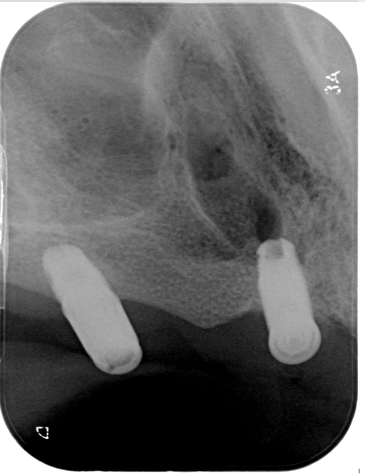
Additional biopsies of the tonsils under general anesthesia reveals HPV mediated p16+ fragments of squamous cell carcinoma in both tonsils (Figure 2).
Therefore a final diagnosis of bilateral synchronous palatine tonsillar carcinoma (cT4aN2M0 on left side and cT1N2M0 on right side). Both lesions were HPV mediated - p16 positive on immunostaining. N2 bilateral cervical nodal disease is also pathologically confirmed.
Differential diagnosis
The differential diagnosis of bilateral palatine tonsil lesions on imaging includes tonsillitis +/- abscess which is usually seen in young adults and associated with fever. There is also an associated intra-peri tonsillar rim enhancing fluid collection. This was highly unlikely in our case. Non Hodgkin’s lymphoma (NHL) which is usually submucosal and bilateral is another differential in our case. However, in NHL non-necrotic cervical nodes are seen and additional nodes may be present elsewhere with presence of systemic symptoms.
Treatment
Upon the completion of the clinical, radiological and pathological staging, the patient is presented and discussed in the multidisciplinary HN tumor board. The final consensus is to proceed with definitive chemoradiation that includes radiation to a dose of 70 Gy in 35 fractions delivered to the bilateral palatine tonsils and any sites of PET positive nodes over approximately 7 weeks and weekly Cisplatin in a dose of 40 mg/m2 as the chemotherapeutic agent.
The patient underwent chemoradiation treatment for 7 weeks.
Discussion
Synchronous bilateral palatine tonsillar SCC is a rare presentation with less than 40 cases described in the English literature, with the first report published in 1971 [6]. Moreover, less than 10 cases of HPV mediated synchronous bilateral palatine tonsillar carcinoma have been reported [7].
Smoking, alcohol and Human Papillomavirus (HPV) infection are known major risk factors implicated in tonsillar carcinoma. However, there is an increasing association between HPV overexpression and synchronous bilateral tonsillar carcinoma. In a review by Joseph AW et al., three models/mechanisms of pathogenesis were described. This included the theory of ‘Field Cancerization’ in which a persistent HPV infection may cause synchronous carcinomas at different anatomical sites, a second mechanism that multiple independent infections from several HPV types infect different areas of squamous epithelium resulting in carcinoma at different sites and a third postulated mechanism that primary HPV-induced tonsillar carcinoma subsequently generated a monoclonal second primary carcinoma in the contralateral tonsil by migration of HPV-infected cells [8,9].
The role of imaging is to provide accurate radiological staging. Several changes have been made to the oropharyngeal cancer staging in the recently released 2018 AJCC Cancer Staging Manual. The most important of which is that the staging of the palatine tonsillar cancers is now dependent on the HPV status of the cancer. This is based on extensive data showing improved outcome in HPV + patients and the need for de-escalation of treatment for these patients [10]. Understanding the key distinctive features involved in the different T and N stages can hone the radiologist’s evaluation of the tumor and its locoregional extent.
MRI is the preferred imaging modality due to its superior soft tissue/tumor contrast [11,12].
Also, in comparison to CT, MR images are less degraded by dental amalgam. This tissue contrast is highlighted by the visualization of the tumor with multiple sequences including T1- weighted (T1), fat-suppressed T2W (T2FS) and post-contrast fatsuppressed (T1+C) images in axial, coronal and sagittal planes. In addition, Diffusion-Weighted (DWI) imaging can be added to further increase the accuracy of lesion characterization. This latter sequence is also useful in the early determination of tumor response to chemotherapy [13]. In cases of tonsillar cancer, the borders of the tumor should be delineated with respect to tonsillar pillars, glossotonsilar sulcus, base of tongue, and the superior constrictor muscle. Invasion of deep/extrinsic muscles of tongue, medial-lateral pterygoid muscles, pre-epiglottic fat, larynx, arterial encasement and prevertebral fascia should be assessed, which are important factors for tumor resectability [14].
Bony invasion (e.g. of mandible, pterygoid plates, hard palate or skull base) can be sought out. Lastly, perineural spread can be suspected with enlargement and abnormal enhancement of the nerves within greater and lesser palatine canal, the pterygopalatine fossa, foramen rotundum, infraorbital fissure/canal, as well as foramen ovale [14,15].
The limitations of MRI include difficulty in visualization of small mucosal & submucosal lesions. The appearance of palatine tonsils is also variable and at times asymmetrical, thus further making the diagnosis of synchronous malignancy challenging. PET-CT is an important adjuvant modality, reinforcing the MR findings with abnormal increased radiotracer uptake in bilateral tonsils.
NCCN guidelines provide no clear recommendation on the treatment of bilateral palatine tonsillar SCC. In a recent report by Christine M Kim et al. they stressed that the contralateral tonsil should routinely be removed in the setting of a known unilateral HPV positive palatine tonsillar SCC since missing a occult contralateral malignancy has grave prognostic implications [9]. Rokkjaer et al. concluded that bilateral tonsillectomy should be recommended in all patients with suspected or biopsy-proven palatine tonsillar SCC (unilateral or bilateral) and in those with carcinoma of unknown primary. However, there is no clear consensus whether bilateral tonsillectomy should be considered as a standard procedure [6,9,16]. Definitive chemoradiation is an acceptable alternate therapy. In these cases, the radiation field must include both tonsils/primary site as well as any sites of positive regional nodes.
Recognition of synchronous malignancy therefore in the contralateral tonsil is essential in minimizing the chance of early recurrence, decreasing morbidity and improving the five year survival rate.
Learning points/take home messages
1. Synchronous bilateral palatine tonsillar SCC is a rare presentation. HPV mediated (p16 positive) synchronous bilateral TC cases are even less commonly reported in the
literature.
2. MRI is integral in imaging work up of these patients and
is indispensable for evaluation and radiological staging. A
systematic MR protocol and review of images is essential.
When combined with PET-CT, the diagnostic accuracy can
further improve.
3. Possibility of a synchronous PCT should be entertained
with equivocal imaging or clinically findings of the contralateral tonsil, especially in those with HPV positive SCC.
Biopsy of the contralateral tonsil can confirm the diagnosis and decrease the possibility of early recurrence.
References
- Carlander AF, Gronhoj Larsen C, Jensen DH, et al. Continuing rise in oropharyngeal cancer in a high HPV prevalence area: A Danish population-based study from 2011 to 2014. Eur J Cancer. 2017; 70: 75-82.
- Marur S, D’Souza G, Westra WH, Forastiere AA. HPV-associated head and neck cancer: A virus-related cancer epidemic. Lancet Oncol. 2010; 11: 781–789.
- Roeser MM, Alon EE, Olsen KD, et al. Synchronous bilateral tonsil squamous cell carcinoma. Laryngoscope. 2010; 120: S181.
- Erkal H, Mendenhall W, Amdur R, et al. Synchronous and metachronous squamous cell carcinomas of the head and neck mucosal sites. J Clin Oncol. 2011; 5: 1358-13625.
Jain KS, Sikora AG, Baxi SS, et al. Synchronous cancers in patients with head and neck cancer: Risks in the era of human papillomavirus-associated oropharyngeal cancer. Cancer. 2013; 10: 1832- 1837.
Rokkjaer MS, Klug TE. Prevalence of synchronous bilateral tonsil squamous cell carcinoma: A retrospective study. Clinical Otolaryngology. 2018; 43: 1–6.
Shimizu F, Okami K, Ebisumoto K, et al. Synchronous HPV-Related Cancer of Bilateral Tonsils Detected Using Transoral Endoscopic Examination with Narrow-Band Imaging. Hindawi Case Reports in Otolaryngology. 2017; 5.
Joseph AW, Ogawa T, Bishop JA, et al. Molecular etiology of second primary tumors in contralateral tonsils of human papillomavirus-associated index tonsillar carcinomas. Oral Oncol. 2013; 49: 244–248.
Kim CM, Maie A. St. John. Should the Contralateral Tonsil Be Removed in Cases of HPV-Positive Squamous Cell Carcinoma of the Tonsil?. Laryngoscope. 129.
Lill C, Bachtiary B, Selzer E. et al. A 5-year update of patients with HPV positive versus negative oropharyngeal cancer after radiochemotherapy in Austria. Wien Klin Wochenschr. 2017; 129: 398–403.
Weber Al et al. Malignant tumors of the oral cavity and oropharynx: clinical, pathologic and radiologic evaluation. Neuroimaging Clin N Am. 2003; 13: 443-464.
Wippold FJ II: Head and neck imaging: The role of CT and MRI, J Magn Reson Imaging. 2007; 25: 453–465.
Schouten CS, Graaf PD, Bloemena E. et al. Quantitative DiffusionWeighted MRI Parameters and Human Papillomavirus Status in Oropharyngeal Squamous Cell Carcinoma American Journal of Neuroradiology. 2015; 36: 763-767.
Yousem DM, Gad K, Tufano RP: Resectability issues with head and neck cancer, AJNR Am J Neuroradiol. 2006; 27: 2024–2036.
Ibrahim M, Parmar H, Gandhi D, et al. Imaging nuances of perineural spread of head and neck malignancies. J Neuro-ophthalmol. 2007; 27: 129–137.
Theodoraki MN, Veit JA, Hoffmann TK et al. Synchronous bilateral tonsil carcinoma: Case presentation and review of the literature. Infect Agent Cancer. 2017; 26: 38-017-0146-5.