
Journal of Clinical Images and Medical Case Reports
ISSN 2766-7820
Case Report - Open Access, Volume 2
A nodule under the toenail
Caroline de Lorenzi*; Sandrine Quenan
Division of Dermatology and Venereology, Geneva University Hospitals, Geneva, Switzerland
*Corresponding Author : Caroline de Lorenzi
Geneva University Hospitals, rue Gabrielle-Perret-Gentil 4, 1205 Genève, Switzerland.
Email: Caroline.deLorenzi@hcuge.ch
Received : Apr 13, 2021
Accepted : May 06, 2021
Published : May 11, 2021
Archived : www.jcimcr.org
Copyright : © Lorenzi CD (2021).
Keywords: Subungual exostosis; Osteocartilaginous tumor.
Citation: Lorenzi CD, Quenan S. A nodule under the toenail. J Clin Images Med Case Rep. 2021; 2(3): 1134.
Description
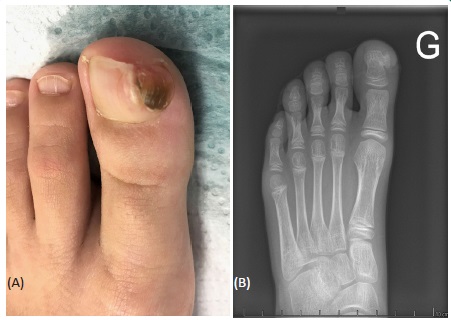
A nine-year-old girl, with no personal or familial medical past history, presented with skin thickness near the left big toenail. It appeared 6 months prior to presentation, initially as a white spot. There is no history of trauma to this site. We found a nodular thickness of the lateral side of the first toenail with axis deviation (Figure 1A). The dermoscopy revealed small thin vessels. The others nails of both feet were normal. We completed with an X-ray (Figure 1B).
What is the diagnosis?
Medial exostosis of the second phalange of the first toe.
Subungual Exostosis (SE) is a rare, benign osteocartilaginous tumor of the distal phalanx [1,2]. It appears as a firm, fixed nodule with a hyperkeratotic surface on the distal phalange of the fingers or toes, especially the great toe [1,2]. The etiology is unknown [1]. Some mecanisms have been suggested to play a role in its development : infection, trauma, activation of a cartilaginous cyst, tumor and hereditary abnormality [1-3]. In some cases, rearrangement in collagen genes COL12A1 and COL4A5 have been found [2]. Differential diagnoses include subungual or periungual fibroma, fibrokeratoma, subungual warts, keratinized pyogenic granuloma, ingrown nail, osteochondroma and less comonly, subungual squamous cell carcinoma or melanoma [1-3].
Diagnosis is confirmed via X-ray imaging: SE appears as exophytic bony growth protrubing from the bone [1,3]. Vascular ectasia, ulceration, hyperkeratosis, onycholysis can be visualized in dermoscopy [1]. Treatment is based on total resection of the exostosis. Recurrences are described in cases of incomplete removal [1-3].
Subungual exostosis is a rare and benign pathology. It should be considererd when confronted with painful subungual lesions.
References
- Khezami M, Abdennadher A, Bellaaj H, Znagui T, Hamdi M, et al. Turrett›s exostoses: About 35 cases. Pan Afr Med J. 2018; 29: 229.
- Das PC, Hassan S, Kumar P. Subungual Exostosis - Clinical, Radiological, and Histological Findings. Indian Dermatol Online J. 2019; 10: 202-203.
- Demirdag HG, Tugrul Ayanoglu B, Akay BN. Dermoscopic features of subungual exostosis. Australas J Dermatol. 2019; 60: e138-e141.