
Journal of Clinical Images and Medical Case Reports
ISSN 2766-7820
Case Report - Open Access, Volume 2
Fracture in a girl with graves’ disease induced by osteoporosis: A case report
Kotb Abbass Metwalley, MD1*; Hekma Saad Farghaly, MD2
1 Professor of Pediatrics, Pediatric Endocrinology Unit, Pediatric Department, Faculty of Medicine, Assiut University, Assiut, Egypt.
2 Professor Pediatrics, Pediatric Department, Faculty of Medicine, Assiut University, Assiut, Egypt.
*Corresponding Author : Kotb Abbass Metwalley
Professor of Pediatrics, Pediatric Endocrinology Unit, Pediatric Department, Faculty of Medicine, Assiut University, Assiut, Egypt.
Email: kotb72@gmail.com
Received : Apr 14, 2021
Accepted : May 10, 2021
Published : May 13, 2021
Archived : www.jcimcr.org
Copyright : © Metwalley KA (2021).
Abstract
A 6-year-old girl with long-standing Graves’ Disease (GD) presented with a left oblique non-displaced humeral fracture. Examination reveals signs of thyrotoxicosis and mild swelling of her left humerus. Investigations confirmed severe overt hyperthyroidism due to GD. A dual-energy X-ray Absorptiometry (DXA) of the lumbar vertebrae (L1-4) and femoral neck revealed that Z-score were -2.9 and -2.1, respectively, representing below the expected range for age which is consistent with osteoporosis. The girl was commenced on carbimazole in a dose of 5 mg two times daily and propranolol 10 mg two times daily. After one year of treatment, she returned to a euthyroid state, and Bone Mineral Density (BMD) increase by 19% and 23% of the lumbar spine and femur neck, respectively
Keywords: Osteoporosis; Graves’ disease; Fracture.
Citation: Metwalley KA, Farghaly HS. Fracture in a girl with graves’ disease induced by osteoporosis: A case report. J Clin Images Med Case Rep. 2021; 2(3): 1142.
Introduction
Pediatric Graves’ Disease (GD) accounts for 10-15% of thyroid disorders in patients less than 18 years of age and has a female predilection [1]. The presentation may be with classical symptoms of thyrotoxicosis, however, hyperactivity, poor school performance, and lack of sleep result in referral to various specialties [2]. Ophthalmopathy and cardiovascular manifestations are rare in childhood GD as compared to adults. Accelerated bone age is common [3]. Osteoporosis is rare in children. When it does occur, it is usually caused by an underlying medical disorder or by drugs [4]. Herein, we reported a 6-year-old girl with long-standing GD who presented with left humeral fracture due to osteoporosis.
Case presentation
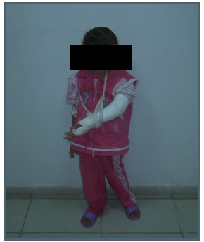
A 6-year-old girl known to have GD since the age of 3 years with very compliance to anti-thyroid mediation and missed follow up appointment, presented to our institution with severe pain and inability to move the left arm following exposure to mild trauma. There was a strong family history of autoimmune thyroid disease, although both parents were unaffected. The evaluation revealed an anxious-looking child with diffuse grade 2 goiter and thyroid eye disease. The pulse rate was 146/min and blood pressure 126/68 mmHg. Heart sounds were normal. His height was 119 cm (>97 centile), and weight was 13 kg (<10 centile). There was mild swelling of her left humerus, with minimal point tenderness over the humerus Figure 1 Examination of other systems were normal. investigations confirmed the clinical picture with a suppressed TSH (<0.1 mU/l) and raised thyroid hormone concentrations, with with a free thyroxine (FT4) of 135.9 pmol/l (normal range 11–23 pmol/l) and free triiodothyronine (FT3) of 40 pmol/l (normal range 3.5–6.5 pmol/l) suggestive of severe overt hyperthyroidism. Neck ultrasonography showed an enlarged heterogeneous echogenic thyroid gland with increased vascularity determined by the color Doppler method. Thyroglobulin and microsomal antibodies were raised at 1/320 and 1/450 respectively. Thyroid receptor antibody was positive (8.9 U/l). Serum calcium of 9.8 mg/dL, phosphorus of 4.8 mg/dL, and alkaline phosphatase of 431 IU/L. Her urine calcium/creatinine was 0.46. Blood gas analysis showed no acidosis (pH 7.36, bicarbonate=22.7) with ultrasound kidneys being normal and creatinine of 0.5 mg/dL. Plasma 25-hydroxy vitamin D was 24 ng/mL (normal=9-37.6 ng/mL) Serum parathyroid hormones were 27.9 pg/mL (normal=12-95 pg/mL). X-ray shoulder showed beside the osteoporotic bone, right oblique non-displaced humeral fracture. The bone age of the child was 7 years. A Dual-energy X-ray Absorptiometry (DXA) of the lumbar vertebrae (L1-4) and femoral neck revealed that Z-score were -2.9 and -2.1, respectively, representing below the expected range for age which is consistent with osteoporosis. The fracture humerus was treated with the hanging arm cast and she was commenced on carbimazole in a dose of 5 mg two times daily and propranolol 10 mg two times daily. After one year of treatment, she returned to a euthyroid state, and BMD increase by 19% and 23% of the lumbar spine and femur neck, respectively.
Discussion
The present case had clinical and biochemical evidence consistent with diagnosis of severe overt hyperthyroidism due to GD. In addition, the fracture of the left humerus after mild trauma was attributed to osteoporosis. Although decreased BMD has been reported at diagnosis of hyperthyroidism in children with GD [5], the fracture itself is a rare presentation in children with GD [6]. Cheruvu et al. [6] reported a 3-year-old boy who presented with a right oblique femur fracture and was found to have GD like our case. Moreover, the patient had a prominent goiter and significant proptosis. Both for our patient and in this case report, low BMD as measured by DXA was consistent with osteoporosis. In adults, excess thyroid hormone is associated with decreased bone mineral density independent of BMI, [7] and history of hyperthyroidism increases the risk for hip fractures, [8] especially at diagnosis.
The receptors for thyroid hormones and TSH are present in bone. These hormones may act directly on both osteoclasts and osteoblasts [7]. Thyroid hormone stimulates bone resorption through a direct effect on osteoclast function, overt hyperthyroidism correlates with accelerated bone turnover and an increased risk of osteoporosis. Moreover, the severity of hyperthyroidism correlates with the decrease in BMD and the increase in fracture risk [9]. TSH suppression in patients with hyperthyroidism might activate osteoclast function, leading also to osteoporosis. Moreover, as GD is associated with weight loss which in turn, is associated with loss of bone mass, it is reasonable to assume that low bone density seen in cases with hyperthyroidism is due to both weight loss and the effect of thyroid function on the bone [10].
The index case showed normalization of the thyroid function tests and improvement of BMD after treatment with antithyroid drugs. This in agreement with Lucidarme et al [5] who prospectively evaluated BMD at diagnosis and 1 and 2 years after initiation of medical treatment in 26 children with GD. They found severe osteopenia at diagnosis which was corrected after 1 and 2 years of treatment to a euthyroid state. This illustrates the importance of thyroid hormone on skeletal growth and bone remodeling [11].
Conclusion
This case emphasized the importance of thyroid hormone on bone remodeling and that neglected and untreated prepubertal children with GD can may result in fracture.
References
- Lavard L, Ranlov I, Perrild H, Andersen O, Jacobsen BB. Incidence of juvenile thyrotoxicosis in Denmark-A nationwide study. Eur J Endocrinol. 1994; 130: 565–568.
- Segni M, Leonardi E, Mazzoncini B, Pucarrelli I, Pasquino AM. Special features of Graves’ disease in early childhood. Thyroid. 1999; 9: 871–877.
- Menon PS, Singh GR. Hyperthyroidism in children: an Indian experience. J Pediatr Endocrinol Metab. 1996; 9: 441–446.
- Rauch F, Plotkin H, DiMeglio L, Engelbert RH, Henderson RC, et al. Fracture prediction and the definition of osteoporosis in children and adolescents: the ISCD 2007 Pediatric Official Positions. J Clin Densitom. 2008; 11: 22.
- Lucidarme N, Ruiz JC, Czernichow P, Léger J. Reduced bone mineral density at diagnosis and bone mineral recovery during treatment in children with Graves’ disease. J Pediatr. 2000; 137: 56–62.
- Cheruvu S, Alverson BK, Quintos JB. Femoral fracture as a rare presentation of prepubertal Graves disease. J Pediatr. 2013; 162: 429–430.
- van der Deure WM, Uitterlinden AG, Hofman A, et al. Effects of serum TSH and FT4 levels and the TSHRAsp727Glu polymorphism on bone: The Rotterdam Study. Clin Endocrinol (Oxf). 2008; 68: 175–181.
- Cummings SR, Nevitt MC, Browner WS, et al. Study of Osteoporotic Fractures Research Group. Risk factors for hip fracture in white women. N Engl J Med. 1995; 332: 767–773.
- Vestergaard P, Mosekilde L. Fractures in patients with hyperthyroidism and hypothyroidism: A nationwide follow-up study in 16, 249 patients. Thyroid. 2002; 12: 411–419.
- Karga H, Papapetrou PD, Korakovouni A. Bone mineral density in hyperthyroidism. Clin Endocrinol (Oxf). 2004; 61: 466-472.
- Goswami R, Shah P, Ammini AC. Thyrotoxicosis with osteomalacia and proximal myopathy. J Postgrad Med. 1993; 39: 89-90.