
Journal of Clinical Images and Medical Case Reports
ISSN 2766-7820
Case Report - Open Access, Volume 2
Chikungunya neonatal: A neglected disease
Suyen Heizer Villela1; Giuliana Villela Pereira2; Maria Elisabeth Lopes Moreira3*
1 Instituto Fernandes Figueira (IFF/ Fiocruz), Brazil.
2 Hospital Universitário Gaffrée e Guinle-UNIRIO, Brazil.
3 Instituto Fernandes Figueira (IFF/Fiocruz), Brazil.
*Corresponding Author: Maria Elisabeth Lopes
Moreira
Instituto Fernandes Figueira (IFF/Fiocruz), Avenida
Rui Barbosa, 716 Flamengo- Rio de Janeiro- RJ, CEP:
22250-020, Brazil.
Email: bebeth@iff.fiocruz.br
Received : May 13, 2021
Accepted : Jun 25, 2021
Published : Jun 30, 2021
Archived : www.jcimcr.org
Copyright : © Moreira MEL (2021).
Abstract
Chikungunya virus infection is an emerging arbovirus with a global distribution that can cause significant morbidity and also death in infected fetuses and neonates. Unfortunately, there is still lack of data about the incidence of Chikungunya in pregnant women and the consequences for their fetus. This is a case series report including clinical presentation, images and clinical assessment.
Keywords: Chikungunya; neonatal; vertical transmission.
Key clinical message: Neonatal Chikungunya by vertical transmission can present with a serious disease with skin lesions and shock and the newborn may need intensive neonatal care. Neonatal disease is more common when the maternal infection occurs close to delivery.
Citation: Villela SH, Pereira GV, Moreira MEL. Chikungunya neonatal: A neglected disease. J Clin Images Med Case Rep. 2021; 2(3): 1210.
Introduction
Chikungunya virus infection is an emerging arbovirus with a global distribution that can cause significant morbidity and also death in infected fetuses and neonates. It is an acute febrile disease caused by the Chikungunya Virus (CHIKV), which is transmitted to humans by the bite of an infected female Aedes aegypti or Aedes albopictus mosquito. CHIKV can affect people of any age or gender, but the signs and symptoms tend to be more intense in children and the elderly [1,2]. The infection is spreading throughout Latin America, causing concern among health authorities about the accelerated spread of a virus that had been previously limited to African and Asian countries [3]. According to the last Epidemiological Bulletin from Brazilian Ministry of Health, the incidence of CHIKV infection in the State of Rio de Janeiro, Brazil, in 2018 was 202.8 cases/100,000 habitants and until September 2019, 447.7 cases/100,000 habitants [4]. Unfortunately, there is still lack of data about the incidence of Chikungunya in pregnant women and the consequences for their fetus. Congenital transmission of arboviruses has serious consequences for neonates [2,5]. In cases of CHIKV, reports suggest that fetuses are more affected when the infection occurs near the birth. The overall pooled risk of symptomatic neonatal disease is 15.5% after maternal infection during gestation. However, the pooled risk is about 50.0% among mothers who are viremic during labour [1,2,5].
Methods
We described two cases of neonatal CHIKV that need a neonatal intensive care. Consent form was obtained from the mothers to describe these cases and about the images use.
Results
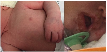
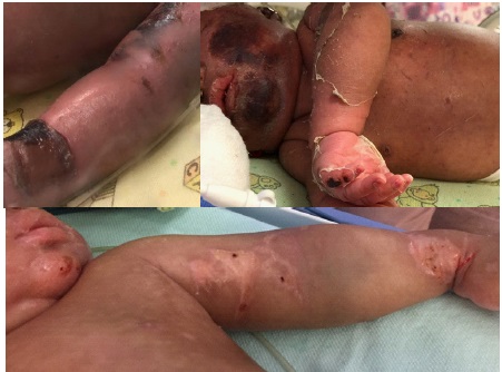
During a Brazilian outbreak in 2018, two cases with positive CHIKV RT-PCR with a similar clinical profile and evolution were admitted to the Neonatal Unit, located in Niteroi City, State of Rio de Janeiro. In both their mothers were symptomatic with fever, joint pain and skin rash near to delivery, the neonates were delivered at term and by c-section, both were exclusively breastfed. Infection by Zika Virus and others serologies were ruled out from them and their mothers. On the 5th day of life, the babies presented with fever, hypoactivity, poor feeding, pain during exam (based on Neonatal Facial Coding System) and cutaneous rash. They evolved with apnea, seizure, peripheral vasodilation, edema, erythroderma, mucosal and skin lesions (Figure 1,2), myocardial dysfunction, thrombocytopenia (minor 12.000 µl), hepatomegaly (with high serum glutamic-oxaloacetic transaminase) and respiratory failure. The babies condition improved gradually. Test results included normal cranial ultrasonography, electroencephalogram, fundoscopy and hearing screening by otoacoustic emissions; ultrasound of the abdomen showed the presence of hepatomegaly, echocardiogram with myocardial dysfunction; negative cultures. In the first case the Magnetic Resonance Imaging of skull had periventricular hyperechogenicity and the second one was normal.
Discussion
Skin manifestations were related by Kumar et al [5] in 25% of cases and correspond to the cutaneous rash, which is usually of the maculopapular type affecting predominantly the thorax, face and limbs, with scaling of the extremities (Figure 1A; B). In the most severe cases erythroderma and edema are observed. In the cases described above, we can observe lesions similar to those found in Stevens-Johnson Syndrome (SJS) and Toxic Epidermal Necrolysis (TEN), which consist of vesicle-bullous, purpuric and necrotic lesions that develop into hyper and or hypopigmentation (Figure 1C). The principal differences between them are the sites (SJS and TEN, all body areas can be involved while CHIKV infection usually extremities, thorax and face) , time of resolution (SJS and TEN may take days to weeks and CHIKV infection usually within 7–10 days), lesions of mucosae (SJS and TEN have severe erosions that heal very slowly while CHIKV infection generally absent but if present, mild erosions that heal in 3 or 4 days) and finally the histopathology [5]. The treatment provided was continuous monitoring, analgesia and wound care for patient comfort, ventilation, hemodynamic and nutritional support, antibiotic therapy and infusion of blood and its derivatives, if necessary.
The descriptions of the cases above, illustrate clinical presentations in the neonatal period, with hospital staying between 32 (Case 1) to 57 days (Case 2). Until now there is no predictive factor of severity in these cases, so that, it is imperative to observe these neonates from the first week of life because CHIKV infection can lead to a serious disease with life threatening complications.
Clinical manifestations among affected neonates were observed within three to seven days after delivery (ranging from 1 to 12 days) and included fever, poor feeding, rash and peripheral edema [5]. In general, these symptoms appears after maternity discharge and because of that all the newborns from mothers presenting CHIKV near delivery need to be monitored.
Author contributions: SHV and GVP describe the cases, request permission to parents to publish and write and approve the draft. MELM review the cases, write, and approve the final text.
References
- Moizéis RNC, Fernandes TAAM, Guedes PMDM, Pereira HWB, Lanza DCF, et al. Chikungunya fever: A threat to global public health. Pathog Glob Health. 2018; 28: 1-13.
- Contopoulos-Ioannidis D, Newman-Lindsay S, Chow C, LaBeaud AD. Mother-to-child transmission of Chikungunya virus: A systematic review and meta-analysis. PLoS Negl Trop Dis. 2018; 12: e0006510.
- Yactayo S, Staples JE, Millot V, Cibrelus L, Ramon-Pardo P. Epidemiology of Chikungunya in the Americas. J Infect Dis. 2016; 214: S441-S445.
- Epidemiological Bulletin from Brazilian Ministry of Health. 2019.
- Kumar S, Agrawal G, Wazir S, Kumar A, Dubey S, et al. Experience of Perinatal and Neonatal Chikungunya Virus (CHIKV) Infection in a Tertiary Care Neonatal Centre during Outbreak in North India in 2016: A Case Series. J Trop Pediatric. 2018.