
Journal of Clinical Images and Medical Case Reports
ISSN 2766-7820
Case Report - Open Access, Volume 2
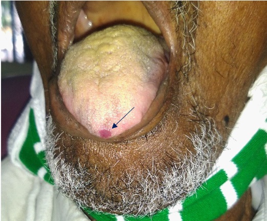
Capillary hemangioma on the tip of the tongue
Karthik R*; Ravikumar PT; Saramma Mathew fenn
Department of Oral Medicine and Radiology, Vinayaka Missions Sankarachariyar Dental College, Vinayaka Missions Research Foundation (Deemed to be University), Salem, Tamilnadu, India.
*Corresponding Author: R Karthik
Department of Oral Medicine and Radiology, Vinayaka Missions Sankarachariyar Dental College, Vinayaka
Missions Research Foundation, Salem, Tamilnadu,
India.
Email: drkarthik@vmsdc.edu.in
Received : May 08, 2021
Accepted : Jul 02, 2021
Published : Aug 03, 2021
Archived : www.jcimcr.org
Copyright : © Karthik R (2021).
Citation: Karthik R, Ravikumar PT, Fenn SM. Capillary hemangioma on the tip of the tongue. J Clin Images Med Case Rep. 2021; 2(4): 1216.
Clinical image description
Hamangioma is a benign, local malformation of blood vessels and grow along with the same rate as the adjacent tissues, they do not invade underlying structures but may involute over a period of time resulting in fine telangiectasia or scarring. Haemangioma can occur anywhere in the body and also in the oral cavity. In tongue they may get ruptured and result in spontaneous bleeding [1].
Capillary Haemangioma are bright red in colour and usually asymptomatic do not cause pain and can be discovered by careful inspection and diascopy test, in which the blanching of the coloured lesion is noted after placing a microscopic glass slide on it. The colour of the lesion appears again once the pressure applied from the microscopic glass slide is removed. The signs and symptoms depend on the location of the haemangioma. The final extent of the lesion can be clearly delineated by MRI (Magnetic Resonance Imaging) [2]. 90 % of the lesions may not be clinically apparent at birth but appear and become more apparent within about 6 months of life. The various treatment modalities include pulsed dioded lasers, KTP laser, radiofrequency ablation, cryotherapy, intralesional injection of Pingyangmycin in concentration of 1mg/ml was found to have cure rate of around 96.4% [3-5].
References
- Gillett D, Fahmy F, Eveson JW, Shotton JC. Intramuscular capillary hamartoma of the tongue. J Laryngol Otol. 2003; 117: 734-735.
- Cappabianca S, Del Vecchio W, Giudice A, Colella G. Vascular malformations of the tongue. MRI findings on three cases. Dentomaxillofac Radiol. 2006; 35: 205-208.
- Mirbehbahani N, Rashidbaghan A. Treatment process for capillary hemangioma. Iran J Ped Hematol Oncol. 2014; 4: 127-130.
- Kutluhan A, Bozdemir K, Uğraş S. The treatment of tongue haemangioma by plasma knife surgery. Singapore Med J. 2008; 49: 312-314.
- Liu XJ, Qing ZP, Li KL. Treatment of hemangioma in oral and maxillofacial region with intralesional injection of pingyangmycin. Shanghai Kou Qiang Yi Xue. 2001; 10: 295-298.