
Journal of Clinical Images and Medical Case Reports
ISSN 2766-7820
Case Report - Open Access, Volume 2
Rare giant zenker’s diverticulum with mediastinal extension
Khadija El Bouhmadi*; Anass Chaouki; Youssef Oukessou; Sami Rouadi; Redallah Abada; Mohamed Roubal; Mohamed Mahtar
Otorhinolaryngology and Head and Neck surgery department, Ibn Rochd University Hospital, Faculty of Medicine and Pharmacy, Hassan II University, Casablanca, Morocco.
*Corresponding Author: Khadija El Bouhmadi
Otorhinolaryngology and Head and Neck surgery
department, Ibn Rochd University Hospital, Faculty of
Medicine and Pharmacy, Hassan II University,
Casablanca, Morocco.
Email: elbkhadija25@gmail.com
Received : Jun 24, 2021
Accepted : Aug 04, 2021
Published : Aug 10, 2021
Archived : www.jcimcr.org
Copyright : © El Bouhmadi K (2021).
Citation: El Bouhmadi K, Chaouki A, Oukessou Y, Rouadi S, Abada R, et al. Rare giant zenker’s diverticulum with mediastinal extension. J Clin Images Med Case Rep. 2021; 2(4): 1259.
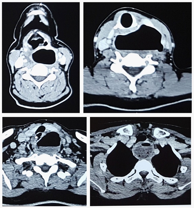
Clinical Image description
Zenker’s Diverticulum (ZD) is a herniation of the posterior pharyngeal wall between the inferior pharyngeal constrictor and the cricopharyngeus muscle through a natural weakness [1]. The main clinical presentation is progressive solid food and pill dysphagia, with subsequent weight loss, halitosis, and regurgitation, complicated by the occurrence of several aspiration and recurrent pneumonia when extended into the chest [2,3]. Indeed, only rare few cases of massive mediastinal extension were reported in the literature [3].
We report the case of 56-year-old woman, with 6 months history of persistent and progressive solid dysphagia, regurgitation and significant weight loss. She also reported chronic coughing and multiple episodes of pneumonia. The physical examination revealed a huge anterior cervical mass, progressively growing, tender and painless at palpation with no signs of local inflammation. The CT scan showed a giant out-pouching sac over the posterior cervical esophagus with air-fluid level extended to its thoracic portion into the mediastinum corresponding to a giant Zenker’s diverticulum. Resection was chosen as a therapeutic option with complete resolution of the symptoms.
References
- Achkar E. Zenker’s diverticulum. Dig Dis. 1998; 16: 144- 151.
- Bock JM, Van Daele DJ, Gupta N, Blumin JH. Management of Zenker’s diverticulum in the endoscopic age: Current practice patterns. Ann Otol Rhinol Laryngol. 2011; 120: 796-806.
- Bock JM, Petronovich JJ, Blumin JH. Massive Zenker diverticulum. Ear Nose Throat J. 2012; 91: 319-320.