
Journal of Clinical Images and Medical Case Reports
ISSN 2766-7820
Case Report - Open Access, Volume 2
Hepatic hemangioma: A case of spontaneous involution
Nida Choudry*; Mohamed Shoreibah
Department of Gastroenterology and Hepatology at University of Alabama, Birmingham, AL, USA.
*Corresponding Author: Nida Choudry
Department of Gastroenterology and Hepatology
at University of Alabama, Birmingham, AL, USA.
Email: nchoudry@uabmc.edu
Received : July 13, 2021
Accepted : Aug 16, 2021
Published : Aug 20, 2021
Archived : www.jcimcr.org
Copyright : © Choudry N (2021).
Citation: Choudry N, Shoreibah M. Hepatic hemangioma: A case of spontaneous involution. J Clin Images Med Case Rep. 2021; 2(4): 1273.
Introduction
Hepatic hemangiomas are benign, well-circumscribed tumors often found incidentally on imaging. They are estimated to be prevalent in 0.4-20% of the population. While usually solitary lesions, these tumors tend to not cause any issues for patients. However, when they grow to be larger than 5 cm, there can be associated symptoms including pain, nausea, or early satiety from compression on adjacent structures. There can be more rare, serious complications including hemobilia, rupture, coagulopathy, or high output heart failure seen in tumors that are typically larger than 10 cm. While small hepatic hemangiomas (<5 cm) that do not cause symptoms do not need to be followed, ones that are greater than 5 cm should be followed at least once to ensure that they are stable in size, or more regularly if they are growing. Symptomatic large hepatic hemangiomas can be intervened on with a number of procedures if needed. These include hepatectomy, enucleation, transcatheter arterial embolization, radiofrequency ablation, and in rare cases, liver transplant.
Case report
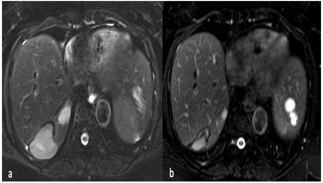
A 66-year-old African American female with a history of hypertension presented to her PCP in 2016 with complaints of nausea. During evaluation she was found to have numerous hepatic cysts and hemangiomas so the patient was referred to hepatology at that time. The largest hemangioma was found to be 7.6 cm in size in the posterior hepatic segment on MRI. Given the patient did not have pressing signs/symptoms, no intervention was done. Between 2018 and 2021, the patient’s hemangioma decreased in size seen on MRI (Figure 1). When the images were originally read by radiology, they were reported as successful partial ablation of segment VII hepatic hemangioma, but after further interdisciplinary review, were determined to be near complete spontaneous involution/thrombosis given lack of history of intervention. The patient continues to be symptom free, and in fact at last visit, denies any further nausea.
Conclusion
This is a rare presentation of spontaneous involution of a hepatic hemangioma seen in a 66-year-old female. Literature regarding this area is scarce so this case aims to highlight this phenomena. The size of hepatic hemangiomas so rarely change this drastically in a short time frame without intervention that the authors thought it was an interesting case to report on.