
Journal of Clinical Images and Medical Case Reports
ISSN 2766-7820
Case Report - Open Access, Volume 2
Ear and pubic keloids in an adolescent girl, the experience of a successful treatment
Harouna Moussa1; Ibrahim Mamadou Abdoul Kadir2*; Sidi Oumoulkhairi1
1 Dermatology Department, Dosso Regional Hospital, Nigeria.
2 Medicine Department, Dosso Regional Hospital, Nigeria.
*Corresponding Author: Ibrahim Mamadou Abdoul
Kadir
Medicine Department, Dosso Regional Hospital, Nigeria.
Email: kader.ibrahim@yahoo.fr
Received : Jun 22, 2021
Accepted : Aug 19, 2021
Published : Aug 23, 2021
Archived : www.jcimcr.org
Copyright : © Kadir IMA (2021).
Keywords: keloid; melanoderma; corticosteroid therapy; recurrence.
Citation: Moussa H, Kadir IMA, Oumoulkhairi S. Ear and pubic keloids in an adolescent girl, the experience of a successful treatment. J Clin Images Med Case Rep. 2021; 2(4): 1279.
Clinical image description
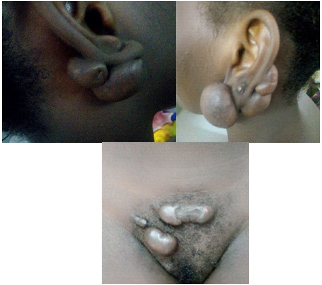
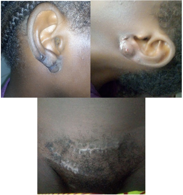
Keloids are frequent connective tissue tumors characterized by a significant proliferation of fibroblasts and collagen with a recurrent and extensive tendency. They can be secondary to a solution of cutaneous continuity: surgical wound; vaccination; burn; hard inflammatory lesion. Sometimes spontaneous, and are more frequently observed in black populations [1-3]. We report the case of a 20 year old women, melanoderma, with no known pathological history who presented to our department with multifocal slightly pruritic tumor lesions evolving for 4 years on the auricle and the pubis. The patient and her companions thought of a mystical disease. The interrogation revealed that these lesions occurred after an ear piercing and folliculitis lesions on the pubic area. The examination revealed a good general condition, firm non sensitive normochromic tumor lesions of variable size with smooth surface, protruding and adherent to the deep plane located on the pubis and bilaterally on the auricle. The CBC and blood glucose levels were normal. We proceeded to a complete removal of these lesions and infiltration sessions of corticosteroids (Triamcinolone) after healing every 2 weeks with periodic control of blood glucose and blood pressure. After 6 infiltration sessions, the result was spectacular, to the great satisfaction of the patient and the Dermatology Department team. No recurrence after one year of follow-up.
References
- Turki IM. Chéloïde géante du bras: Traitement médico-chirurgical.
- Philandrianos C, et al. Les cicatrices chéloïdes (première partie) : une pathologie de la cicatrisation cutanée. Ann Chir Plast Esthet. 2015.
- Kelly T, Huang Y, Simms AE, Mazur A. Chapter Three - Fibroblast Activation Protein-α: A Key Modulator of the Microenvironment in Multiple Pathologies. International Review of Cell and Molecular Biology. 2012; 297: 83-116.