
Journal of Clinical Images and Medical Case Reports
ISSN 2766-7820
Case Report - Open Access, Volume 2
Neglected microphone in the ear: About a case
Sefrioui Taha Ismail1*; Ait Taleb Hajar1; Boumendil Ikram1; Nitassi Sophia2; Bencheikh Razika2; Oujilal Abdelilah2; Benbouzid Mohamed Anas2; Essakalli Leila2
1 Resident Physician, Department of Otorhinolaryngology and Head & Neck surgery, IbnSina University Hospital, Morocco.
2 Professor, Department of Otorhinolaryngology and Head & Neck Surgery, IbnSina University Hospital, Mohammed V University, Rabat, Morocco.
*Corresponding Author: Sefrioui Taha Ismail
Resident Physician, Department of Otorhinolaryngology and Head & Neck Surgery, IbnSina university
Hospital, Morocco.
Email: taha.ismail.sefrioui@gmail.com
Received : Jun 18, 2021
Accepted : Aug 25, 2021
Published : Aug 31, 2021
Archived : www.jcimcr.org
Copyright : © Ismail ST (2021).
Abstract
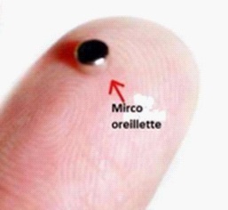
Stranger bodies (EC) of the middle ear are among the emergencies least encountered by otolaryngologists (ENT). A 16-year-old patient with no otological history had seen a right chronic otorrhee with otalgia and hypoacousia for 6 months. Microscopic examination of the diseased ear had resulted in purulent secretions with inflammation of the external auditory canal. After aspiration, a blackish appearance was observed occupying the lower quadrants of the eardrum with associated myringitis. The gentle mobilization with the suction cannula and a spike revealed a fixed, painful CE, impacting the tympanic membrane and deeply introduced into the middle ear. The extraction of the EC could be carried out without difficulty (Figure 1), completed by myringoplasty with an aponevrotic graft. The first observation of a Tallic CE of the middle ear has been neglige for more than 20 years reported to you in 1883 by Lucius Holland [4]. Since then, few cases have been written down. Syms and Nelson reported 4 cases of EC in the middle ear with chronic otitis media [2]. Average EC penetrants from the ear are relatively rare. They may go unnoticed, especially in children, to reveal themselves later by complications. Hence the interest of prevention and diagnosis precoce allowing a simple extraction.
Keywords: foreign body; extraction; middle ear.
Citation: Ismail ST, Hajar AT, Ikram B, Sophia N, Razika B, et al. Neglected microphone in the ear: About a case . J Clin Images Med Case Rep. 2021; 2(4): 1285.
Introduction
Stranger bodies (EC) of the middle ear are among the emergencies least encountered by otolaryngologists (ENT). They account for 1% of otological conditions and less than 0.4% of all consultations with emergency services [1]. Voluntary selfadministration in the ear usually occurs in children [2]. If the body moving from the middle ear is known to me, it can lead to a significant morbidity, especially if it is tall and if it is not managed before co-management [3]. We report the observation of a young girl who had consulted for a persistent otorrh and whose body was found in the middle ear.
Observation
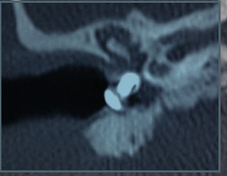
A 16-year-old patient with no otological history had seen a right chronic otorrh with otalgia and hypoacousia. This otorrhea had been growing for 6 months, not at all, of average abundance, from clench to bathing and slightly improved by topical and general dication treatment taken in self dication without prealable otoscopy. It was accompanied by hypoacousia and otalgia, recently appearing with paroxysmal episodes of vertiginous sensation, without otorragy or facial paralysis associated. Microscopic examination of the diseased ear had resulted in purulent secretions with inflammation of the external auditory canal. After aspiration, a blackish appearance was observed occupying the lower quadrants of the eardrum with associated myringitis. The gentle mobilization with the suction cannula and a spike revealed a fixed, painful CE, impacting the tympanic membrane and deeply introduced into the middle ear. The sign of fistula was negative and vestibular and neurological examination without abnormality. The notion of introducing a CE in the ear was later found in the interview, as the young person had inserted it for 6 months, which had caused a passage otalgie and otorragie. The audiogram showed a so-called straight transmission with an average Rinne of 40 dB. The Computed Tomography (CT) of rocks had objective an EC of 12 mm in length and height density, occupying the bottom of the external auditory canal up to the eardrum crate in contact with the promontory with a total liquid-like filling of the eardrum crate and the posterior cavities The surgical exploration of the eardrum crate was carried out under general anesthesia by a first post route, because the EC was impacted in the eardrum, was drawn in the middle ear, and had caused problems. It had the presence of a rearranged metal nail, strongly enclosed in the tympanum, in contact with the promontory and surrounded by fibrosis. The lining of the crate was polyploıde and inflammatory. The ossicular chain was intact and continuous without contact with the foreign body. The facial nerve canal was intact and there was no labyrinthine fistula. The extraction of the EC could be carried out without difficulty (Figure 2), completed by myringoplasty with an aponevrotic graft. Two months later, the patient no longer complained of any symptoms and the otoscopy of covered an intact neotympan. The test audiogram was normal.
Discussion
The first observation of a Tallic CE of the middle ear has been neglige for more than 20 years reported to you in 1883 by Lucius Holland [4]. Since then, few cases have been written down. Syms and Nelson reported 4 cases of EC in the middle ear with chronic otitis media [2]. The EC known to the middle ear, either intentionally or accidentally, are rare, their frequency may be estimated to be less than 0.4% in the different litter ages [3]. A predominance of the age range between 2 and 15 years has been observed by the different authors. Boys appear to be more affected than girls (56%) with favourable socio-economic conditions [5]. The main clinical manifestations related to the introduction of a CE in the middle ear are: otalgia, the sensation of a double-dipping of the ear, hypoacousia, otorragia, acouphe and vertigo. These ECs may also be affected by an infectious symptomatology with otorrhea and may be prolonged, or incidentally, during a routine otoscopic examination. They may, however, go unfit and return to the stage of complications such as otitis externa, masto ıdite, chronic otitis media, labyrinthitis or neurological complication [6]. Panosian and Dutcher reported a case of facial nerve zion by a metastatic EC which is not transferred to a welder [7]. Another case of facial paralysis with hypoacousia has also been reported by Sce`ne and Vinding from an EC in the middle ear [7]. In our observation, the EC was affected by a persistent otorrh and hypoacousia linked to an open ear infection. When the EC is present in the middle ear, there is the problem of the inside of the retro-tympanic ments: ossicles, facial nerve, cochle and vestibule. The assessment should include an audiogram and a scanner of the rocks [7]. In our observation, the audiogram was used to document the hypoacousia and its type, and CT to assess the nature, exact location and relationship of the EC to the middle ear. The discovery of a CE imposes its urgent extraction under microscope, sometimes under general anesthesia, especially for children restless uncooperative. The choice of technique depends on the shape, size of the CE and especially the position and duration of his stay in the ear [8]. It can also be removed in consultation with a speculum. If the tympanic perforation does not close within 6 weeks, a myringoplasty must be programmed. A complete surgical extraction by myringoplasty, rarely used, is indicated in complicated forms and in cases of impossibility of extraction under a microscope [9].
In our observation, the EC was very much impacted in the eardrum and, localized in the middle ear, it had caused lesions, which had motivated an exploration of the eardrum body by a way initially posterior. Postoperative evolution is often favorable in the absence of attainment of the noble elements of the middle ear.
Conclusion
Average EC penetrants from the ear are relatively rare. They may go unnoticed, especially in children, to reveal themselves later by complications. Hence the interest of prevention and diagnosis precoce allowing a simple extraction.
References
- Das SK. Anetiological evaluation of foreign bodies in the ear and nose. J Laryngol Otol. 1984; 98: 989–91.
- Syms 3rdC, Nelson RA. Impression-material foreign bodies of the middle ear and external auditory canal. Otolaryngol Head Neck Surg. 1998; 119: 406–7.
- Schimanski G. Silicone foreign body in the middle ear caused by auditory canal impression in hearing aid fitting. HNO.1992; 40: 67–8.
- Dubois M, Franc¸ oisM, Hamrioui R. Corpse´trangers del’oreille; A` propos d’unese´rie de40cas. Arch Pediatr. 1998; 5: 970–3.
- Supiyaphun P, Sukumanpaiboon P. Acuteotalgia: A case report of mature termite in the middle ear. Auris NasusL arynx. 2000; 27: 77–8.
- Eleftheriadou A, Chalastras T, Kyrmizakis D, etal. Metallic foreign body in middle ear: An unusual cause of hearing loss. Head Face Med. 2007; 3: 23.
- Panosian M,Dutcher J. Trans tympanic facial nerve injury in welders. Occup Med. 1994; 44: 99–101.
- Lin CC, HoK Y, Wang LF, etal. Foreign-body-induced otitis media: A casereport. Kaohsiung. J Med Sci. 2002; 18: 585–8.
- Jacob A, Morris TJ, Welling DB. Leaving alasting impression: Ear mold impressions as middle ear foreign bodies. Ann Otol Rhinol Laryngol. 2006; 115: 912–6.