
Journal of Clinical Images and Medical Case Reports
ISSN 2766-7820
Case Report - Open Access, Volume 2
Role of endoscopic ultrasonography in the diagnosis of dysphagia aortica
Ramin Niknam; Gholam Reza Sivandzadeh*
Gastroenterohepatology Research Center, Shiraz University of Medical Sciences, Shiraz, Iran.
*Corresponding Author: Gholam Reza Sivandzadeh
Gastroenterohepatology Research Center (GEHRC),
Shiraz University of Medical Sciences, Namazi Hospital,
Third floor, 71935-1311 Shiraz, Iran.
Email: ghsivand@sums.ac.ir
Received : Aug 01, 2021
Accepted : Sep 03, 2021
Published : Sep 08, 2021
Archived : www.jcimcr.org
Copyright : © Sivandzadeh GR (2021).
Abstract
Dysphagia aortica is an unusual cause of dysphagia. Diagnosis of this condition is usually based on chest radiography, barium esophagogram, chest computed tomography, endoscopy, and aortography. We report a case of dysphagia aortica who was successfully diagnosed using endoscopic ultrasonography and treated with life style modification and medications.
Keywords: dysphagia aortica; aortic aneurysm; endoscopic ultrasonography; endoscopy.
Citation: Niknam R, Sivandzadeh GR. Role of endoscopic ultrasonography in the diagnosis of dysphagia aortica. J Clin Images Med Case Rep. 2021; 2(5): 1302.
Introduction
Dysphagia refers to problems with the passage of swallowed material from the mouth to the hypopharynx or esophagus. Dysphagia can be caused by a number of benign and malignant disorders. Upper Gastrointestinal (GI) endoscopy is recommended for most of the patients to establish a diagnosis, and when appropriate to perform therapeutic modalities. Early barium swallow evaluation for the patients with esophageal dysphagia is useful. In some cases, esophageal manometry may be necessary [1]. The term of Dysphagia Aortica (DA) refers to dysphagia caused by esophageal compression by a tortuous or aneurysmal aorta [2].
Case report
A 45-year-old woman presented to our center with intermittent non-progressive solid dysphagia from 6 months prior to evaluation. She did not have any anorexia and weight loss. She had poor control high blood pressure for 12 years. Her physical examination findings were normal.
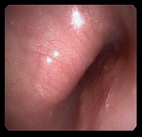
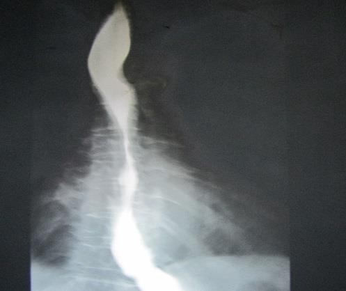
An upper GI endoscopy was performed which revealed extrinsic compression of the esophagus caused by a mass effect at 23 cm from incisors teeth (Figure 1). A barium esophagogram result showed extrinsic compression of the esophagus (Figure 2).
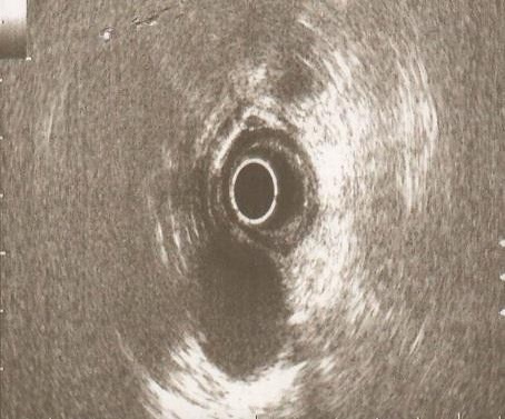
For detection of the possible causes of esophageal compression, endoscopic ultrasonography (EUS) was performed which showed a mass effect on the esophagus at 23 cm of the incisors by aortic arch. by enlarged aortic arch. The sub-mucosal layers of esophagus were intact (Figure 3).
Because of the mild intermittent dysphagia without any evidence of alarming signs and symptoms, lifestyle modification plus drug therapy of hypertension was advised for her under closed follow up. Dysphagia was significantly decreased in follow-up at 12 months after treatment.
Discussion
DA is an unusual cause of dysphagia. Some of cases of dysphagia aortica were reported in literature. Ideal diagnostic and therapeutic modality for these patients is controversial [3-6]. Diagnosis of DA is usually made by history, chest radiography, barium esophagogram, chest Computed Tomography (CT), endoscopy, and aortography [2-6].
Chest CT is useful radiological modality in diagnosis of dysphagia in some patients. CT is used for evaluating both the aortic wall morphology and extra-aortic structures as possible causes of dysphagia [7]. The finding of DA on chest radiography is an unfolded aortic arch below which lies a tortuous dilated aorta. The findings of this condition on barium swallow are partial stricture or flattened contour of the left margin of the esophagus or pulsatile movement of the barium synchronous with aortic pulsation. DA findings on upper GI endoscopy are stenosis or pulsatile compression or kinking of the esophagus, although vascular pulsation is often seen on endoscopic evaluation of the normal esophagus [2].
Several therapeutic modalities have been introduced based on the severity of symptoms of DA including lifestyle modification, treatment of any associated congested heart failure or hypertension, and surgical procedures. Many surgical procedures have been described for the treatment of severe DA [2].
Beachley MC et al, reported a case of DA diagnosed by chest radiograph, barium esophagogram, angiography, and endoscopy and treated by surgery, while initial presentation was long term history of odynophagia [6]. Kim JH et al reported a patient of DA with progressive dysphagia and weight loss diagnosed by chest radiograph, chest CT, and barium esophagogram. The patient declined any invasive procedures [3]. Majumder B et al reported a case of Marfan syndrome with DA diagnosed by chest radiography, chest computed tomography, and aortography, while initial presentation was intermittent chest pain and backache with dysphagia [4]. Hiller HG et al reported an old patient with severe DA and weight loss diagnosed by chest radiograph and chest computed tomography. The patient died because of delayed diagnosis and treatment modalities [5]. Maeder M et al reported a case of aberrant right subclavian artery, anomaly of the aortic arch, diagnosed by using EUS. They concluded that EUS is a simple and excellent tool for the diagnosis of this condition [8].
EUS as a minimally invasive endoscopic modality has become an important tool for the management of various GI and non-GI disorders in many centers. The clinical indications of EUS as an adjunctive endoscopic imaging study for lesions of the GI tract and surrounding organs are varied including assessment of mediastinal disease. EUS has an advantage of low complication rate [9-11].
The choice and effect of treatment of DA patients depends on the baseline symptom severity. The patients with mild symptoms can be treated by avoiding solid foodstuffs likely get stuck in the Esophagus. The therapy of the associated hypertension (low-sodium diet with or without medications) may reduce symptoms. The patients who are not responding to non-invasive therapy may need to esophageal dilatation or surgical procedures [2].
Our unusual case highlights a patient with dysphagia aortic which was suspected by barium esophagography and endoscopy, confirmed by using EUS. Because of her mild intermittent symptoms, the patient was advised to change her diet and drug therapy was started for the management of hypertension. In conclusion, EUS as a simple and minimally invasive endoscopic modality can be performed for the diagnosis of patients with suspected dysphagia aortica and usually does not require confirmation by using other investigation methods.
Conflict of interest: Authors declared that there is no conflict of interests.
References
- Spechler SJ. AGA technical review on treatment of patients with dysphagia caused by benign disorders of the distal esophagus. Gastroenterology. 1999; 117: 233-54.
- Wilkinson JM, Euinton HA, Smith LF, Bull MJ, Thorpe JA. Diagnostic dilemmas in dysphagia aortica. Eur J Cardiothorac Surg. 1997; 11: 222-7.
- Kim JH, Jang SW, Kim DB, Lee HJ, Kim JG, Kwon BJ, Cho EJ, Rho TH, Kim JH. A Patient With Dysphagia due to an Aortic Aneurysm. Korean Circ J. 2009; 39: 258-60.
- Majumder B, Kumar A, Ranjan P, Mondal S. Dysphagia Aortica. Indian Heart J. 2008; 60: 588–590.
- Hiller HG, Lagattolla NR. Thoracic aortic aneurysm presenting with dysphagia: A fatal delay in diagnosis. Thorac Surg Sci. 2007; 4: 1.
- Beachley MC, Siconolfi EP, Madoff HR, Chaudhry RM. Dysphagia aortica. Dig Dis Sci. 1980; 25: 807-10.
- Lee J. Radiological Imaging of Aortic Aneurysms. Korean Circulation J. 2007; 37: 337-347.
- Maeder M, Binek J. Impact of endoscopic ultrasonography in the diagnosis of aberrant right subclavian artery: a case report.[abstract] Ultraschall Med. 2004; 25: 296-8.
- ASGE Training Committee, DiMaio CJ, Mishra G, McHenry L, Adler DG, Coyle WJ, Dua K, DeGregorio B, Enestvedt BK, Lee LS, Mullady DK, Pais SA, Rajan E, Sedlack RE, Tierney WM, Faulx AL. EUS core curriculum. GastrointestEndosc. 2012; 76: 476-81.
- Mesenas S, Ang TL, Khor C, Vu C. Guidelines for endoscopic ultrasonography. Ann Acad Med Singapore. 2010; 39: 489-92.
- ASGE Standards of Practice Committee, Gan SI, Rajan E, Adler DG, Baron TH, Anderson MA, Cash BD, Davila RE, Dominitz JA, Harrison ME 3rd, Ikenberry SO, Lichtenstein D, Qureshi W, Shen B, Zuckerman M, Fanelli RD, Lee KK, Van Guilder T. Role of EUS. Gastrointest Endosc. 2007; 66: 425-34.