
Journal of Clinical Images and Medical Case Reports
ISSN 2766-7820
Case Report - Open Access, Volume 2
Pleomorphic adenoma of the palate
Naourez Kolsi* ; Emna Bergaoui; Rachida Bouatay; Jamel Koubaa
Department of Otorhinolaryngology, Head and Neck Surgery at “Fattouma Bourguiba” Hospital, Monastir, Tunisia.
*Corresponding Author: Naourez Kolsi
Department of Otorhinolaryngology, Head and Neck
Surgery at “Fattouma Bourguiba” Hospital, Monastir,
Tunisia.
Email: kolsi.naourez@yahoo.fr & Kolsi.naourez@gmail.com
Received : Aug 05, 2021
Accepted : Sep 10, 2021
Published : Sep 17, 2021
Archived : www.jcimcr.org
Copyright : © Kolsi N (2021).
Keywords: pleomorphic adenoma; palate; surgical excision.
Citation: Kolsi N, Bergaoui E, Bouatay R, Koubaa J. Pleomorphic adenoma of the palate. J Clin Images Med Case Rep. 2021; 2(5): 1316.
Case presentation
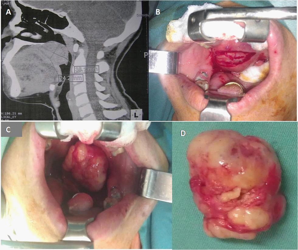
A woman 43 years, presented with 9 years history of a painless swelling in the palatal region, slow-growing. Her medical history was not significant. On intraoral examination, a median ovoid mass measuring 4 cm diameter was found at the junction of hard and soft palate. The mass was firm, with smooth surface. No radiographic evidence of bone involvement was seen on the CT scan (Figure A).
Surgical excision under general anesthesia: excision of the mass was carried out, the overlying mucosa was healthy, so conserved (Figure B,C&D). Histopathologic report confirmed the lesion to be “pleomorphic adenoma”. The lesion has not recurred after four years follow-up.
Final diagnosis: Pleomorphic adenoma of the palate.
Comments
Pleomorphic Adenoma (PA) can be defined as a benign mixed tumor composed of epithelial and myoepithelial cells arranged with fibrous capsule. Palate is the most common site affected of the minor salivary glands. PA may occur at any age, but mainly they affect patients in the fourth and fifth decades, 60% of them are female [1].
Some cases of palatal PA have been published in the literature. The first cases of PA were published in 1957. They reviewed, over a period of 30 years, the tumors of salivary gland origin in patients at 18 years or younger and they noted only two cases of PA of the Palate in over 470 cases of benign salivary gland tumors. One was a recurrent tumor which has first been treated 5 years earlier when the patient was 9 years old. The second case was made with no recurrence [2]
The diagnosis of PA is evoked on the basis of history, physical examination, cytology and confirmed by histopathology.
Clinically palatal pleomorphic adenoma presents as a slowgrowing, unilateral firm mass that may become large if is untreated. In most cases, it occurs on the soft and hard palate due to the highest concentration of salivary glands there and is typically a firm or rubbery submucosal mass without ulceration [3-5].
CT scan and MRI can provide information on the location and size of the tumor and extension to surrounding superficial and deep structures. Fine-needle aspiration cytology can help in the diagnosis.
Three differential diagnosis
- Odontogenic cysts
- Soft tissue tumors such as fibroma, , neurofibroma
- Malignant tumor of minor salivary glands: Cystic adenoid carcinoma.
The treatment consists on a surgical excision with removal of the palatal bone, if involved. Wide excision with negative margins is the optimal strategy for the management of pleomorphic adenomas due to occasional lack of encapsulation, with lower risk of recurrence [3].
A close follow-up is necessary to detect recurrences, which can appear years after the occurrence of the primary lesion.
References
- Ogesh Sharma, Anisha Maria, and Amit Chhabria. Pleomorphic adenoma of the palate. Natl J Maxillofac Surg.2011; 2: 169-171.
- Courten, T. Lombardi, J. Samson: Pleomorphic adenoma of the palate in a child: 9-year follow-up. Int. J. Oral Maxillofac. Surg. 1996; 25: 293-295.
- Mansur Rahnama, Urszula Orzędała-Koszel, Łukasz Czupkałło, Michał Łobacz. Pleomorphic adenoma of the palate: A case report and review of the literature. Wspolczesna Onkol. 2013; 17: 103–106
- Vellios F, Shafer WG. Tumors of minor salivary glands. Surg Gynecol Obstet. 1959; 108: 450-6.
- Ledesma-Montes C, Garces-Ortiz M. Salivary gland tumours in a Mexican sample. A retrospective study. Med Oral. 2002; 7: 324- 30.