
Journal of Clinical Images and Medical Case Reports
ISSN 2766-7820
Case Report - Open Access, Volume 2
Devil is in the detail, subtle signs of posterior reversible encephalopathy syndrome
A Boix Lago1; N Nersesyan2
1 Department of Neurology, Hospital Universitari Doctor Josep Trueta, Girona, Spain.
2 Department of Neuroradiology, Hospital Universitari Doctor Josep Trueta, Girona, Spain.
*Corresponding Author: A Boix Lago
Department of Neurology, Hospital Universitari
Doctor Josep Trueta, Girona, Spain.
Email: almudenaboix.girona.ics@gencat.cat
Received : Aug 18, 2021
Accepted : Sep 14, 2021
Published : Sep 21, 2021
Archived : www.jcimcr.org
Copyright : © Boix Lago A (2021).
Keywords: posterior reversible encephalopathy syndrome; seizures; hypertensive encephalopathy
Citation: Boix Lago A, Nersesyan N. Devil is in the detail, subtle signs of posterior reversible encephalopathy syndrome. J Clin Images Med Case Rep. 2021; 2(5): 1322.
Clinical image description
A 33-year-old woman presented to the hospital with convulsive seizure and hypertensive crisis. As differential diagnosis cortical venous thrombosis, ischaemic or haemorrhagic stroke, space occupying lesion, Posterior Reversible Encephalopathy Syndrome (PRES) and epileptic syndrome were considered. Antiepileptic and antihypertensives drugs were used as initial clinical management.
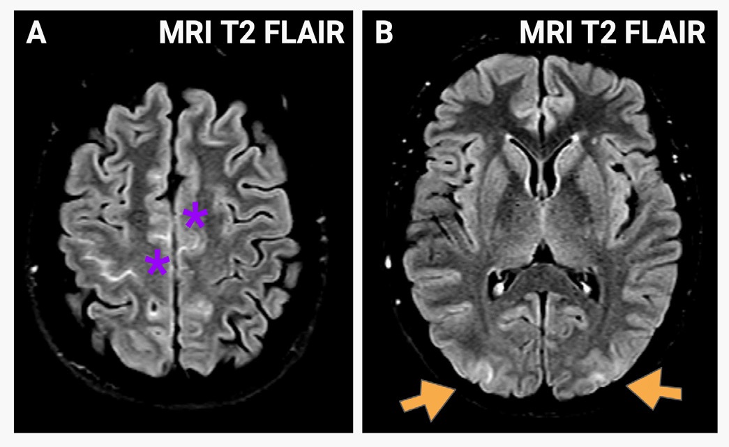
Magnetic Resonance Imaging (MRI) findings in this case include hyperintensity of frontoparietal parasagittal margins and right central sulcus, signs of subarachnoid hemorrhage (A, asterisk, Figure) associated with bilateral and symmetrical increased signal in occipital and parietal region with posterior distribution (B, arrows, Figure) and adequate permeability of the intracranial vascular structures. The neuroimaging along with the clinical context guides the diagnosis.
PRES was first described in 1996 as a wide variety of neurological symptoms along with typical neuroimaging findings. Clinical features include headache, visual symptoms, seizures, focal neurological signs and altered consciousness that usually are reversible [1]. It’s a neurotoxic state that occurs as a result of cerebrovascular dysregulation due to a variety of triggers, primary and secondary causes of hypertension, cytotoxic substances, renal disease, infection and autoimmune disorders [2]. The syndrome is usually visualized in neuroimaging as vasogenic edema predominantly in parieto-occipital region; it may be associated with laminar necrosis and bleeding [3]. In most cases, the prognosis is favorable and symptoms are reversible; although early detection is necessary
Always consider PRES in the case of an hypertensive emergency with neurological symptoms. The fundamental treatment is the control of blood pressure and withdrawal of the cytotoxic drug, if any.
References
- Marlene Fischer, Erich Schmutzhard. Posterior reversible encephalopathy syndrome. J Neurol. 2017; 264: 1608–1616.
- Anderson RC, Patel V, Sheikh-Bahaei N, Liu CSJ, Rajamohan AG, et al. Posterior Reversible Encephalopathy Syndrome (PRES): Pathophysiology and Neuro-Imaging. Front. Neurol. 2020; 11: 463.
- Sarbu et al. White Matter Diseases with Radiologic-Pathologic Correlation. Radio Graphics. 2016.