
Journal of Clinical Images and Medical Case Reports
ISSN 2766-7820
Case Report - Open Access, Volume 2
Bilateral renal infarcts: A mysterious cause of a rare condition
Elhussein AE Elhassan1,2*; Peter J Conlon1,2
1 Department of Nephrology & Transplantation, Beaumont Hospital, Dublin, Ireland.
2 Department of Medicine, Royal College of Surgeons, Dublin, Ireland.
*Corresponding Author: Elhussein AE Elhassan
Department of Nephrology & Transplantation,
Beaumont Hospital, Beaumont Road, 1297, Dublin 9,
Ireland.
Email: elhusseinelhassan@beaumont.ie
Received : Sep 09, 2021
Accepted : Oct 08, 2021
Published : Oct 15, 2021
Archived : www.jcimcr.org
Copyright : © Elhassan EAE (2021).
Abstract
Renal infarction is an uncommon condition. While work up to rule out common causes is sensible, a significant proportion may have no cause detected therefore classified as idiopathic renal infarction. We report a rare cause of renal infarction with initial workup results were of no avail.
Citation: Elhassan EAE, Conlon PJ. Bilateral renal infarcts: A mysterious cause of a rare condition. J Clin Images Med Case Rep. 2021; 2(5): 1366.
Introduction
Renal infarction, resultant from interruption of renal arteries blood flow, is a rare condition. Symptoms vary considerably and Computer Tomography (CT) is considered a gold standard for establishing the diagnosis. We report a mysterious cause of renal infarcts in a candidate of living-kidney donor.
Case report
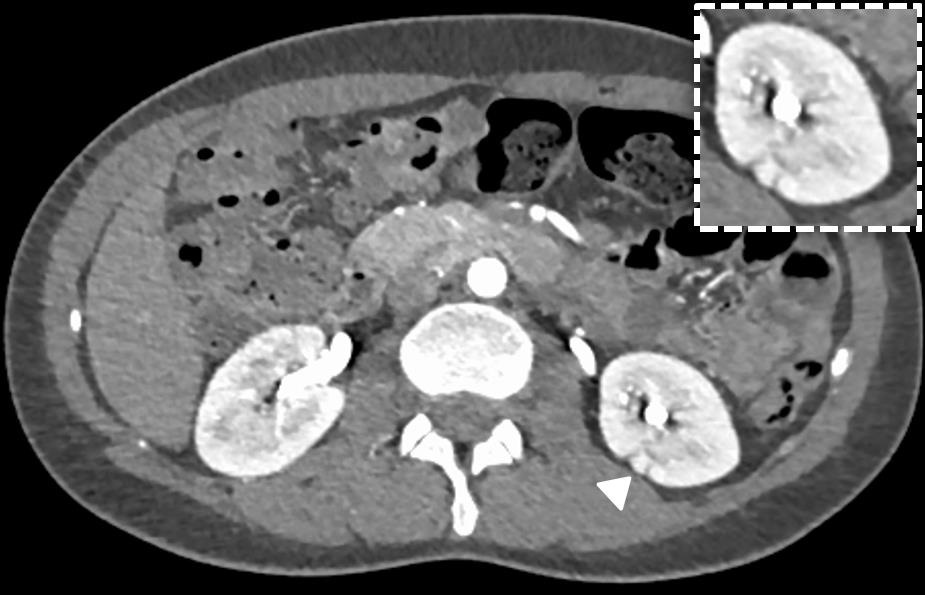
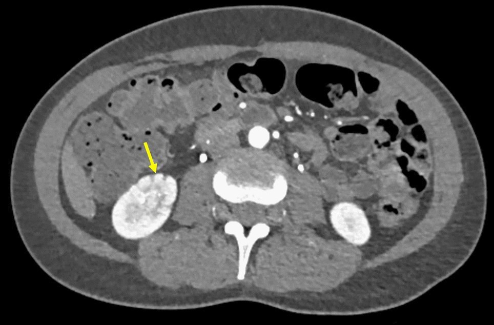
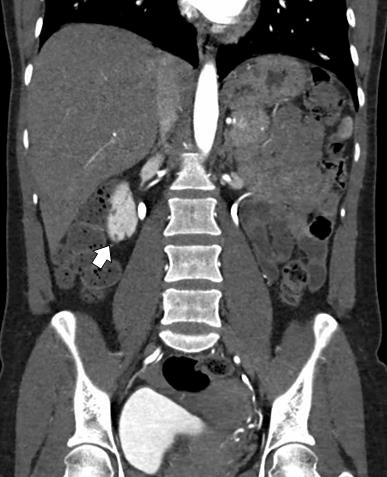
A 46-year-old healthy woman presented for evaluation as a potential kidney donor for her husband. The patient’s history was notable of menorrhagia due to uterine fibroids, which was treated with an uncomplicated uterine artery embolisation about five years ago. Otherwise, she had no significant history of pregnancy-associated complications, vasculitis, thrombosis, or ischemic risk factors. Her renal function was normal, with a creatinine level of 57 μmol per liter (normal range, 45 to 84 μmol per liter) with a normalized technetium 99 GFR clearance of 106 mL/min/1.73 sqm. As part of the evaluation, renal ultrasound showed that the right and left kidneys measure 9.2 cm and 8.5 cm in length, respectively. Subsequently, Computed Tomography Angiogram (CTA) was performed and demonstrated adequately normal renal vasculature with calcified fibroids present in the fundus of the uterus. However, it revealed multiple hypodense areas representing small infarcts in both kidneys but more marked on the right side (Figure 1,2&3). These multiple infarcts in the kidneys were suggestive of prior embolic events. Echocardiogram, holter monitor, autoimmune and vasculitis workup, and hypercoagulable screening were all done and normal. Renal infarcts were thought to be from inadvertent distal embolic showers during Uterine Artery Embolisation (UAE), yet off-target distal embolisation to adjacent organs is one of uncommon reported complications. In fact, it may have long-term consequences; as in this case, she was deemed unsuitable for kidney donation and advised to continue her regular annual follow up with her GP.
Discussion
Here we report a rare finding of bilateral renal infarcts in a potential kidney donor with preserved renal function, which were thought to be from an unintentional remote embolisation of the microparticles to the renal vasculature during uterine artery embolisation (UAE). While UAE is a safe and effective procedure, distal embolisation to other organs has been reported [1,2]. Other sources of embolisation were excluded by a normal cardiac echocardiogram and holter monitor. Similarly, the patient also had negative autoimmune, vasculitic tests as well as hypercoagulable workup.
According to the literature, at the time of the event renal infarction frequently present with persistent abdominal and/or flank pain, but spectrum of non-specific symptoms, like nausea, vomiting, and fever, can occur, mimicking other medical conditions which add extra diagnostic challenges [3,4]. With atrial fibrillation being the commonest cause of renal infarction, cardio-embolic, renal artery disease, hypercoagulable conditions and post procedural complications accounted for the majority of identified aetiologies. Hence, high index of suspicion is required as well as a thorough medical history, including remote procedures. The sensitivity and the high-yield of contrast enhanced CT have made it the gold standard imaging for establishing diagnosis of renal infarctions, in addition to it is relatively non-invasive. In a large retrospective study, CT scans solely were able to diagnose more than 95% of patients with renal infarcts in this cohort [5].
Renal infarction has potentially serious consequences requiring immediate actions to mitigate such harmful sequel. Treatment options, depends on the underlying aetiology, patient clinical status and available expertise, range from administration of heparin, warfarin, antiplatelet treatment, and curative thrombectomy. Examining renal infarction outcomes in a cohort exceeding 400 patients, Oh et al showed that almost one-third of the patients had renal dysfunction, acute kidney injury, newonset chronic kidney disease and end-stage kidney disease in 20.9%, 10.9%, and 2.1%, respectively. Twenty-one patients (5%) died during a median duration of follow up of 20 months [5]. Other studies reported similar results in terms of renal outcomes and mortality rates [4,6].
Conclusion
Renal infarction is a rare condition but potentially harmful. Diagnosis requires a high index of suspicion, in addition to the contrast-enhanced CT scan as the gold-standard imaging modality. Despite aggressive treatment, renal infarction can result in renal dysfunction and increases mortality rates. It is a crucial diagnosis with subsequent sequelae particularly in kidney donors such as our patient.
References
- Vatutin NT, Taradin GG, Smyrnova GS, Valery B. Kostogryz B. Kostogryz, et al. The Successful Treatment of Pulmonary Embolism as a Complication after Uterine Fibroid Embolization (Case Report)”, Current Women`s Health Reviews. 2019; 15: 150.
- Hamoda H, Tait P, Edmonds DK. Fatal pulmonary embolus after uterine artery fibroid embolisation. Cardiovascular and interventional radiology. 2009; 32: 1080–1082.
- Domanovits H, Paulis M, Nikfardjam M, et al. Acute renal infarction. Clinical characteristics of 17 patients. Medicine. 1999; 78: 386-394.
- Marie B, et al. Acute renal infarction: a case series. Clinical journal of the American Society of Nephrology : CJASN. 2013; 8: 392- 8.
- Oh YK, Yang CW, Kim YL, Kang SW, Park CW, Kim YS, Lee EY, Han BG, Lee SH, Kim SH, Lee H, Lim CS. Clinical Characteristics and Outcomes of Renal Infarction. Am J Kidney Dis. 2016; 67: 243- 50.
- Bae EJ. Hwang K Jang HN, et al. A retrospective study of shortand long-term effects on renal function after acute renal infarction. Ren Fail. 2014; 36: 1385-1389.