
Journal of Clinical Images and Medical Case Reports
ISSN 2766-7820
Case Report - Open Access, Volume 2
A rare glans cavernous hemangioma in young patient: A case report
Liu Chunguang1; Bhushan Sandeep2; Xu Xuejun3*
1 Graduate School of Zunyi Medical University, Zunyi, Guizhou 563003, Chengdu Second People’s Hospital, Chengdu, Sichuan 610017, China.
2 Department of Surgery, Chengdu Second People’s Hospital, Chengdu, Sichuan 610017, China.
3 Department of Neurosurgery, Zunyi Medical University, Chengdu Second People’s Hospital, Chengdu, Sichuan 610017, China.
*Corresponding Author: Xu Xuejun
Department of Neurosurgery, Zunyi Medical University,
Chengdu Second People’s Hospital, Chengdu, Sichuan
610017, China.
Email: 809860761@qq.com
Received : Sep 14, 2021
Accepted : Oct 19, 2021
Published : Oct 26, 2021
Archived : www.jcimcr.org
Copyright : © Xuejun X (2021).
Abstract
Background: Cavernous hemangioma of the glans penis is a relatively rare disease in clinical practice, caused by congenital dysplasia.
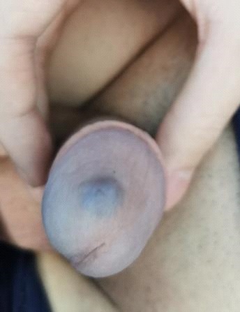
Case presentation: A 20-year-old male, found a snake shaped dark purple mass on the glans penis for more than 10 years. During the operation, the tumor was seen to be about 0.5 cm away from the outer urethra. The tumor was broken longitudinally, and the boundary between the tumor and the corpus cavernosum was not clear, and the tumor was bluntly separated until it was completely removed. Postoperative pathological examination revealed a cavernous hemangioma. The patient was discharged without complications such as infection or wound dehiscence.
Conclusion: With the increase of sexual demand and to avoid rupture and bleeding of glans penis cavernous hemangioma are recommend early intervention and treatment.
Keywords: glans; cavernous hemangioma; treatment methods.
Citation: Chunguang L, Sandeep B, Xuejun X. A rare glans cavernous hemangioma in young patient: A case report. J Clin Images Med Case Rep. 2021; 2(5): 1382.
Background
Hemangiomas are benign tumors or vascular malformations composed of differentiated and mature blood vessels, usually covering only a single layer of endothelium. According to the size of the vascular diameter of hemangioma, the morphology and characteristic histological structure of endothelial cells are divided into five categories: Sponge type, capillary type, vein type, epithelioid type and granulation tissue type [1]. The most common sites for hemangioma are subcutaneous, and can also occur in deep soft tissues and internal organs. Hepatic hemangioma is the most common in internal organs. Tumors of the vascular tissue of the kidney are not uncommon, but they occur in urine [2]. Hemangiomas of the pathway system are rare, with an incidence of only 2%. Sponge and capillary hemangioma are the most common but penile hemangioma is a rare congenital abnormality [3].
Case presentation
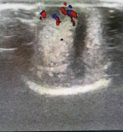
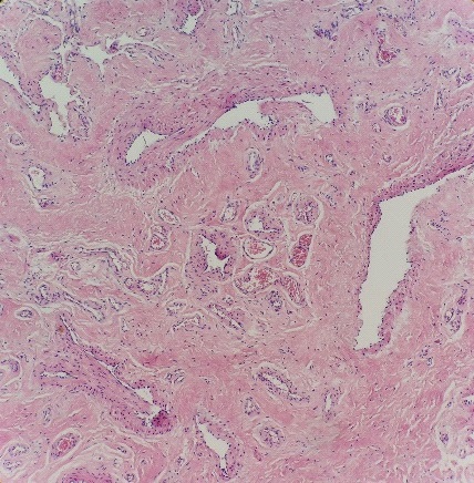
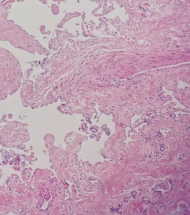
A 20-year-old male, found a snake shaped dark purple mass on the glans penis for more than 10 years (Figure 1). When pressed, the color of the mass is slightly lighter. The patient had no pain, pruritus, redness, swelling, secretion, hematuria, dysuria and weight loss. He had a healthy and asexual life in the past and had no history of smoking, drinking, accidental injury and so on. His physical examination shows the prepuce is too long, the glans can be upturned and exposed, the glans near the urethral orifice is dark purple snake shaped uplift, the size is 1 × 1 cm, tough, no tenderness, clear boundary, protruding on the surface of the glans, no redness, swelling and ulceration; his bilateral inguinal lymph nodes were not palpable. Auxiliary examination shows blood routine, urine routine, liver and kidney function, coagulation items, AIDS, syphilis, hepatitis and other laboratory tests were normal. Color Doppler ultrasound of glans mass showed that hypoechoic mass with a range of 8.5 × 6.5 × 7.0 mm could be seen subcutaneously, the boundary was not clear, the shape was regular, and the internal echo was not uniform and linear blood flow signals could be seen by pressure detection. Ultrasound examination shows a hemangioma (Figure 2). During the operation, the tumor was seen to be about 0.5 cm away from the outer urethra. The tumor was broken longitudinally, and the boundary between the tumor and the corpus cavernosum was not clear, and the tumor was bluntly separated until it was completely removed. Surgeons used absorbable thread for suturing the wound base and forming surface tissue and to avoid the damaging of urethra during the operation. Postoperative pathological examination revealed a cavernous hemangioma (Figure 3 & 4). The patient was discharged after the surgery without any complication of infection and wound dehiscence. Three months later during follow up examination the patient has a normal sexual intercourse life without any complication.
Discussion
Hemangioma is a benign tumor or vascular malformation composed of well differentiated blood vessels. It is usually lined with a single layer of endothelium. According to the caliber of hemangioma, the morphology and characteristic histological structure of endothelial cells can be divided into five types: sponge type, capillary type, venous type, epithelioid type and granulation tissue type. The most common site of hemangioma is skin and subcutaneous, and it can also occur in deep soft tissue and internal organs. Among internal organs, hepatic hemangioma is the most common, vascular tissue tumor in kidney is not rare, and hemangioma in urinary system is rare, accounting for only 2% [4]. Cavernous and capillary hemangiomas are the most common. Penile hemangioma is a rare congenital dysplasia. The commonly used methods for the treatment of glans hemangioma include laser therapy, sclerotherapy, surgical resection, cryotherapy. Laser treatment: laser has deep penetration rate, good coagulation, no fibrosis, less scar formation, but it may need multiple operations, and the cost of treatment is high [5]. The laser therapy reported in the literature includes potassium thiophosphate laser, neodymium: YAG laser (Neodymium: YAG laser). Sclerotherapy: Injection of sclerosing agent into the area of hemangioma can promote the formation of intravascular thrombosis and achieve the effect of fibrosis or sclerosis. The treatment cost is low, and complications such as local skin necrosis, ulcer and pigmentation are prone to occur. The sclerotherapy reported in the literature includes sodium tetradecyl sulfate, 30% hypertonic saline and 2% polydocanol [6]. Surgical treatment: radical resection of the hemangioma was performed by incision and suture. Compared with non operation, it has higher risk of bleeding and scar formation, even erectile dysfunction. Cryotherapy uses the physical properties of refrigeration materials to reduce the skin temperature of local pathological tissue, contract capillaries, necrosis and shedding of tissue cells. It has the advantages of simple operation, no bleeding and low infection rate. The refrigeration material is mainly liquid nitrogen. Other treatments for hemangioma include oral propranolol, oral or injection of corticosteroids, electric cautery. In this case, the location of hemangioma was superficial, the lesion was small, and did not affect the urethra. Combined with the opinions of the patients and their families, the treatment of hemangioma resection and glans reconstruction was adopted.
For asymptomatic hemangioma, most lesions do not need treatment. Some patients care about beauty and function and choose the above treatment. For genital hemangioma, more attention should be paid to the importance of sexual function.
Yong f et al. [7] reported two cases of multiple treatments with hematuria, and finally diagnosed urethral cavernous hemangioma. At the same time, it also warned that sexual life may lead to rupture and bleeding of the tumor. Liu Guizhong [8] also reported that rupture of posterior urethral hemangioma is the main cause of severe hematuria after sex. For such patients, in order to avoid bleeding after sex, we suggest early intervention.
Conclusion
Penile hemangioma is a rare congenital abnormality. With the increase of sexual demand and to avoid rupture and bleeding of glans penis cavernous hemangioma are recommend early intervention and treatment.
Declarations
Ethics approval and consent to participate: The protocol was approved by the Chengdu Second People’s Hospital Clinical Research Ethics Committee, and parents of all subjects provided informed consent.
Consent for publication: Written consent to publish this information was obtained from study participants.
Availability of data and materials: Not applicable, Please contact author for data requests.
Competing interests: The authors declare that they have no competing interests.
Funding: Not applicable
Author contributions:
LC1: First author, Concept/Design, Data analysis/interpretation, drafting article, Critical revision of article, Approval of article, Statistics, Funding, Data collection.
SB1: Concept/Design, Data analysis/interpretation, drafting article, Critical revision of article, Approval of article, Statistics, Funding, Data collection
XX*: Corresponding author, Concept/Design, Data analysis/ interpretation, drafting article, Critical revision of article, Approval of article, Statistics, Funding, Data collection.
Acknowledgements: All authors contribute equally to this manuscript.
References
- Illescas Megias V, Saez Barranquero F, Rojo Cormonal E et al. Hemangioma of the glans penis [J]. Archivos espanoles de urologia. 2014; 67: 357-9.
- Ulker V, Esen T. Hemangioma of the glans penis treated with Nd:YAG laser [J]. International urology and nephrology. 2005; 37: 95-6.
- Mondal S, Biswal D K, Pal D K. Cavernous hemangioma of the glans penis [J]. Urology annals. 2015; 7: 399-401.
- Kim K S, Lee H, Hwang J H, et al. Incidentally detected cavernous hemangioma of the glans penis after circumcision: A case report [J]. Medicine. 2020; 99: e20217.
- Sakhiya J, Sakhiya JD, Virmani N, et al. A Challenging Case Of Cavernous Hemangioma Of The Face Treated With The Combination Of Cryotherapy, Laser, And Dermatosurgery [J]. The Journal of clinical and aesthetic dermatology. 2020; 13: 14-6.
- Kagami S, Kuyano Y, Shibata S, et al. Propranolol is more effective than pulsed dye laser and cryosurgery for infantile hemangiomas [J]. European journal of pediatrics. 2013; 172: 1521-6.
- Yong F, Juan L, Jinhuan W, et al. Urethral cavernous hemangioma: a highly misdiagnosed disease (a case report of two patients and literature review) [J]. BMC urology. 2019; 19: 13.
- Liu Guizhong, Hu Haibing, Wu Baojun, et al. Etiology and treatment of severe hematuria after male sexual life% J Chinese Journal of Urology [J]. 2020; 10: 1.