
Journal of Clinical Images and Medical Case Reports
ISSN 2766-7820
Case Report - Open Access, Volume 2
Catheter knotting in a neonate
Rong Huang; Jia-ying Chen; Ying Zhang; Chang-mei Chen; Jia-liang Zhou*
Department of Neonatal Surgery, Guangdong Women and Children Hospital, Xingnan Road 521, Guangzhou 511400, China.
*Corresponding Author: Jia-liang Zhou
Guangdong Women and Children Hospital, Xingnan
Road 521, Guangzhou 511400, China.
Email: 124171458@qq.com
Received : Sep 27, 2021
Accepted : Oct 26, 2021
Published : Nov 02, 2021
Archived : www.jcimcr.org
Copyright : © Zhou J-L (2021).
Keywords: catheter knotting; complication.
Citation: Huang R, Chen JY, Zhang Y, Chen CM, Zhou JL. Catheter knotting in a neonate. J Clin Images Med Case Rep. 2021; 2(6): 1393.
Clinical image description
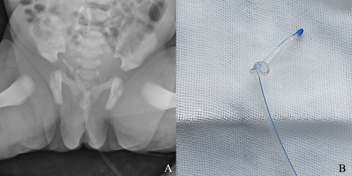
A 36-week preterm neonate with anal atresia and rectoperineal fistula was catheterized with a size 6F Foley’s catheter when she underwent anoplasty on the second day after birth. When the patient recovered from anesthesia, it was decided to remove the catheter. While the catheter was being removed, resistance was felt halfway through the procedure. Abdominal plain films revealed a catheter coiled in the pelvis. With copious lubricant injected into the bladder through the catheter and patient sedated, it was removed by manipulation alone using gentle traction (Figure 1). The infant had no bleeding at the urethral meatus and no obvious abnormality in urination during the 3-month of follow-up.
Catheter knotting is an extremely rare complication of catheterization. The widely accepted hypothesis is that the catheter forms a loop and coils on itself when excessive length of the highly flexible catheter is inserted into the bladder. As the bladder decompresses the catheter tip loops through the coil. It mainly occurs in young boys who have been catheterized with a feeding tube. To our knowledge, this is the first case of Foley’s catherter knotting in a newborn in the literature [1]. The catheter’s relatively wide and tough head increases the difficulty and risk of pulling out the catheter directly. However, it was unexpectedly removed by manipulation alone.
Various methods have been described to remove a knotted catheter such as manipulation alone using gentle traction, manipulation with detangling using a guidewire, manipulation with dilatation, endourology and laparoscopy techniques [1]. From our own exprience, we recommend more attempt at manipulation alone using gentle traction when the uretheral is sufficiently lubricated and patient adequately sedated or anesthetized, especially if the patient is a female, because the female urethra is wider, shorter and more malleable compared with the male.
References
- Singh VP, Sinha S. Spontaneous knotting of urinary catheters placed with nonindwelling intent: Case series and literature review. Urology annals. 2019; 11: 443-46.