
Journal of Clinical Images and Medical Case Reports
ISSN 2766-7820
Case Report - Open Access, Volume 2
Presumed topography of basal ganglion: Derived from TRODAT images in a case of hemorrhagic stroke complicated with asymmetrical cogwheel rigidity
Li-Yin Chen1; Yu-Li Chiu2,6; Yuan-Ling Chang1,3,4; Ruei-Sian Ding1; Shin-Tsu Chang4,5*
1 Department of Medical Education and Research, Kaohsiung Veterans General Hospital, Kaohsiung, Taiwan.
2 Department of Nuclear Medicine, Kaohsiung Veterans General Hospital, Kaohsiung, Taiwan.
3 Department of Anaesthesiology, Kaohsiung Armed Forces General Hospital, Kaohsiung, Taiwan.
4 Department of Physical Medicine and Rehabilitation, Tri-Service General Hospital, School of Medicine, National Defence Medical Center, Taipei, Taiwan.
5 Department of Physical Medicine and Rehabilitation, Kaohsiung Veterans General Hospital, Kaohsiung, Taiwan.
6 Department of Medical Imaging and Radiology, Shu-Zen Junior College of Medicine and Management, Kaohsiung, Taiwan.
*Corresponding Author: Shin-Tsu Chang
Department of Physical Medicine and Rehabilitation,
Tri-Service General Hospital, School of Medicine,
National Defence Medical Center, Kaohsiung Veterans
General Hospital, 386, Dazhong 1st Rd, Zuoying Dist,
Kaoshiung 813414, Taiwan.
Email: ccdivlaser1959@gmail.com
Received : Sep 23, 2021
Accepted : Nov 10, 2021
Published : Nov 17, 2021
Archived : www.jcimcr.org
Copyright : © Shin-Tsu C (2021).
Abstract
Introduction: Systemic maps of basal ganglion lesion corresponding to clinical symptoms are lacking at present. Only the framework of functional domains in striatum was presumed. We present a case with asymmetrical cogwheel rigidity which is related to the lesion site of the basal ganglion in his functional brain image.
Case presentations: A 50-year-old male who suffered from subarachnoid hemorrhage and intracranial hemorrhage presented with upper limbs cogwheel rigidity. The symptom was more severe in the right side than the left side. Dopamine transport images revealed bilateral decreased dopamine transporter binding capacity in bilateral striatum. In left striatum, decreased uptake in dorsal region is more severe than ventral region. Cogwheel rigidity was mildly improved after use of pramipexole.
Conclusions: This case report suggests that asymmetrical cogwheel rigidity is linked to the lesion site of the basal ganglion. Topography exists in the striatum and is related to dysfunctional site of parkinsonism.
Keywords: cogwheel rigidity; asymmetrical cogwheel rigidity; parkinsonism; basal ganglion; topography
Citation: Li-Yin C, Yu-Li C, Yuan-Ling C, Ruei-Sian D, Shin-Tsu C. Presumed topography of basal ganglion: Derived from TRODAT images in a case of hemorrhagic stroke complicated with asymmetrical cogwheel rigidity. J Clin Images Med Case Rep. 2021; 2(6): 1411.
Introduction
Multiple division ways of the Basal Ganglia (BG) had been presumed. The boundaries of functional domain and more detailed subdivision remain unclear. BG dysfunction leads to parkinsonism, which features tremor, bradykinesia, rigidity, and postural instability. The symptoms are thought to be contralateral to the lesion in BG, systemic maps of lesion corresponding to clinical symptoms are lacking at present. We report a case of a male patient with cogwheel rigidity after Subarachnoid Hemorrhage (SAH) and Intracranial Hemorrhage (ICH) and his functional image.
Case report
This is a 50-year-old left-handed male with past history of hypertension. Sudden onset of conscious disturbance with vomiting episode took place on the 1st of March, 2021. Brain Computed Tomography (CT) and brain Computed Tomography Angiography (CTA) revealed diffuse SAH with Intraventricular Hemorrhage (IVH) and acute hydrocephalus, anterior communicating artery aneurysm rupture related. He received emergency surgery of external ventricular drainage placement for decompression. Seizure episode lasting for ten minutes had been noted once. Progressive brain edema with delayed intracerebral hematoma and midline shift was found on following brain CT on the 6th of March. Emergency right frontal-temporal craniotomy with removal of ICH was performed. Following brain CT on the 15th of March revealed decreased intracerebral hematoma volume. Left ventriculoperitoneal shunt and right cranioplasty were performed on the 18th of March. Management did not improve the level of consciousness in this patient. Tracheostomy was then performed. After weaning from mechanical ventilation successfully, he was transferred to rehabilitation ward for rehabilitation.
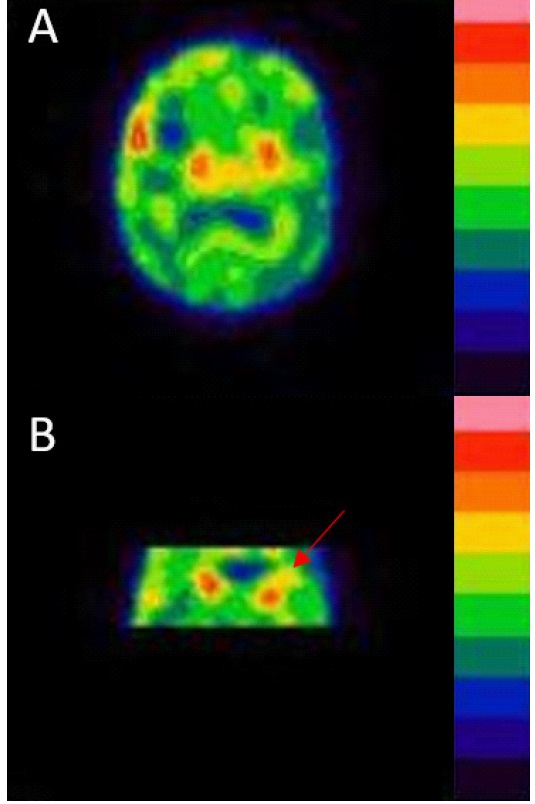
The patient was in totally dependent status and at Brunnstrom stage II. Cogwheel rigidity over bilateral upper extremities persisted with more severe in the right elbow. Tc-99m TRODAT-1 Single-Photon Emission Computed Tomography (SPECT) was arranged and revealed decreased dopamine transporter binding capacity in bilateral striatum. Visual score of striatum was 3 on both sides, which demonstrated decreased caudate uptake with no uptake in putamen (Figure 1). Uptake ratio of striatum is 0.12 and 0.23 on right side and left side. Cogwheel rigidity was mildly improved after use of pramipexole, an aminothiazole dopamine agonist.
Discussion
We used Tc-99m TRODAT-1 SPECT to evaluate topographic relationship between cogwheel rigidity and BG in our case. The result revealed decreased uptake in bilateral sides of caudate together with no uptake in putamen, mimicking the result of Parkinson’s disease (PD) patient. In left striatum, decreased uptake in dorsal region was more severe than ventral region. On the other hand, ventral region of right striatum showed fewer uptakes than dorsal region (Figure 1).
Clinically, cogwheel rigidity affects the right upper limb more than the left upper limb in this patient. Symptom is thought to be contralateral to lesion in BG. Dorsal striatum is mainly connected to premotor and motor cortex [1]. Therefore, more severe decreased uptake in left dorsal region corresponds to cogwheel rigidity severity. Another aspect of asymmetrical cogwheel rigidity may be related to injury of SAH and ICH. Followup CT of this patient showed right cerebrum atrophy. Less cogwheel rigidity in left upper limb is probably related to ongoing recovery of abnormal muscle tone.
Comprehensive anatomical maps of striatum are lacking while compared to the cortex and thalamus. Based on neuroanatomy of animal, striatum of mice is mainly comprised of three known functional domains. Sensorimotor domain and associative domain are located in the dorsolateral striatum and the dorsomedial striatum respectively [2]. Ventral striatum is thought to be a component of the emotional system which can control motivated behavior [3]. Hunnicutt et al. declared a fourth functional domain, in the posterior region of striatum, which has more input from distinct areas of the cortex and thalamus [4,5]. Yamamoto et al. presumed that posterior end striatum played a role in visual-motor skills[6]. Connectivity patterns of human BG and cortex were studied by magnetic resonance diffusion weighted imaging data. Segregation and overlapping of input and output coexist in human BG. Probabilistic functional tractography in human is presented and corresponds to animal anatomic test [1].. While the framework of functional domains in striatum has been provided. More detailed domains related to parkinsonism need to be studied. Alberto Cacciola et al. tried to explain the detailed pathophysiology of movement disorders by cortico-BG networks [7]. The precise pathophysiology of BG structure leading to parkinsonism needs to be investigated.
Dopamine transporter binding capacity in BG can be evaluated by Tc-99m TRODAT-1 SPECT. It has been used for diagnosing and evaluating PD recently [8,9].Visual score can reflect neurodegeneration and is used to evaluating severity of disease [10]. Tc-99m TRODAT-1 SPECT is also helpful for studying the relation ship between BG dysfunction and various movement disorders [11,12]. Further detailed pathophysiology could be evaluated based on this functional image.
Dopamine agonist has been proved to have anti-parkinsonism effect by activating dopamine receptors, and several studies have demonstrated dopamine agonist can be used as monotherapy of PD [13,14]. Pramipexole, non-ergoline dopamine agonist, has the superior efficacy than bromocriptine, due to its affinity for different dopamine receptors[15]. Pramipexole has dopaminergic side-effects, such as nausea, somnolence and hypotension, but its side effect is less common than ergoline dopamine agonist[16]. No side effect of pramipexole was found in our patient. Mild improvement of cogwheel rigidity was found.
Conclusion
Only the framework of functional domains in striatum was presumed, while detailed systemic functional map of BG remains uncertain. We used Tc-99m TRODAT-1 SPECT to evaluate functional capacity of different regions in the striatum. Combining images and clinical symptoms, we interpreted that topography existed in the striatum and was related to dysfunctional site of parkinsonism.
References
- Draganski B, Kherif F, Klöppel S, Cook PA, Alexander DC, Parker GJ, Deichmann R, Ashburner J, Frackowiak RS. Evidence for segregated and integrative connectivity patterns in the human Basal Ganglia. J Neurosci. 2008; 28: 7143-52.
- Yin HH, Knowlton BJ. The role of the basal ganglia in habit formation. Nat Rev Neurosci. 2006; 7: 464-76.
- Gruber AJ, McDonald RJ. Context, emotion, and the strategic pursuit of goals: interactions among multiple brain systems controlling motivated behavior. Front Behav Neurosci. 2012; 6: 50.
- Hunnicutt BJ, Long BR, Kusefoglu D, Gertz KJ, Zhong H, Mao T. A comprehensive thalamocortical projection map at the mesoscopic level. Nat Neurosci. 2014; 17: 1276-85.
- Hunnicutt BJ, Jongbloets BC, Birdsong WT, Gertz KJ, Zhong H, Mao T. A comprehensive excitatory input map of the striatum reveals novel functional organization. Elife. 2016; 5: e19103.
- Yamamoto S, Kim HF, Hikosaka O. Reward value-contingent changes of visual responses in the primate caudate tail associated with a visuomotor skill. J Neurosci. 2013; 33: 11227-38.
- Weng YH, Yen TC, Chen MC, Kao PF, Tzen KY, Chen RS, Wey SP, Ting G, Lu CS. Sensitivity and specificity of 99mTc-TRODAT-1 SPECT imaging in differentiating patients with idiopathic Parkinson’s disease from healthy subjects. J Nucl Med. 2004; 45: 393- 401.
- Cacciola A, Milardi D, Bertino S, Basile GA, Calamuneri A, Chillemi G, Rizzo G, Anastasi G, Quartarone A. Structural connectivity-based topography of the human globus pallidus: Implications for therapeutic targeting in movement disorders. Mov Disord. 2019; 34: 987-996.
- Huang WS, Lin SZ, Lin JC, Wey SP, Ting G, Liu RS. Evaluation of early-stage Parkinson’s disease with 99mTc-TRODAT-1 imaging. J Nucl Med. 2001; 42: 1303-8.
- Huang WS, Lee MS, Lin JC, Chen CY, Yang YW, Lin SZ, Wey SP. Usefulness of brain 99mTc-TRODAT-1 SPET for the evaluation of Parkinson’s disease. Eur J Nucl Med Mol Imaging. 2004; 31: 155- 61.
- Ho YJ, He HC, He YK, Chang ST. Appearance of lower limb symptom in a case of prodromal parkinsonism during the postpartum period. World Journal of Physical and Rehabilitation Medicine 2019; 3: 1013.
- Hung CJ, Wang SC, Cheng YY, Chang ST. Brain imaging findings in Parkinson disease with Pisa syndrome: A case report. Medicine (Baltimore). 2021; 100: e24631.
- Clarke CE, Guttman M. Dopamine agonist monotherapy in Parkinson’s disease. Lancet. 2002; 360: 1767-9.
- Schwarz J. Rationale for dopamine agonist use as monotherapy in Parkinson’s disease. Curr Opin Neurol. 2003; 16: S27-33.
- Brooks DJ. Dopamine agonists: their role in the treatment of Parkinson’s disease. J Neurol Neurosurg Psychiatry. 2000; 68: 685-9.
- Contin M, Lopane G, Mohamed S, Calandra-Buonaura G, Capellari S, De Massis P, Nassetti S, Perrone A, Riva R, Sambati L, Scaglione C, Cortelli P. Clinical pharmacokinetics of pramipexole, ropinirole and rotigotine in patients with Parkinson’s disease. Parkinsonism Relat Disord. 2019; 61: 111-117.