
Journal of Clinical Images and Medical Case Reports
ISSN 2766-7820
Case Report - Open Access, Volume 2
Dead man sign of ECG: An unique predicting sign of acute coronary syndrome
Mayank Yadav
PGIMER, Advanced Cardiac Center, Sector 12 Chandigarh 160012, India.
*Corresponding Author: Mayank Yadav
PGIMER, Advanced Cardiac Center, Sector 12
Chandigarh 160012, India.
Email: mayank001.my@gmail.com
Received : Sep 30, 2021
Accepted : Nov 12, 2021
Published : Nov 19, 2021
Archived : www.jcimcr.org
Copyright : © Yadav M (2021).
Abstract
Coronary artery disease is the most common cause of morbidity and mortality worldwide. Acute coronary syndrome which includes STEMI, NSTEMI and unstable angina commonly diagnosed with the help of 12 lead in ECG in ER with or without elevated biomarkers. Inferior wall myocardial infarction is common cause of ST elevation myocardial infarction with low mortality rate.
In this case report, we present a 52 years old male presented to ER with the complain of typical chest pain for more than 2 hours duration. Pain was in center of chest with radiation to back and left arm and associated with excessive sweating. Patient is a known smoker for past 10 years. At presentation Blood pressure was 110/70 mmHg and pulse rate of 55/min. Troponin I level was five times of upper normal limit. Basic investigations including ECG were done at presentation. ECG was showing typical changes of inferior wall myocardial infarction with infarction of right ventricle with hidden unique sign known as dead man sign commonly predict the location of obstruction and course of disease in the setting of acute coronary syndrome.
Citation: Yadav M. Dead man sign of ECG - An unique predicting sign of acute coronary syndrome. J Clin Images Med Case Rep. 2021; 2(6): 1415.
Introduction
This case represents inferior wall MI with unique dead man sign which predict the culprit artery and level of the block. This sign is commonly associated with the course of bradyarrhythmias and tachyarrhythmias.
Inferior wall of heart receives it blood supply from posterior descending artery branch of right coronary artery in about 70% people and in 10% it is the branch of left circumflex artery in rest 20% it receives dual blood supply form both RCA and LCX [1].
Inferior wall MI consists of 40% of all acute myocardial infarction and occurs commonly due to acute obstruction of right coronary artery with mortality rate less than 10%. Diagnosis of inferior wall infarction is made by 12 lead ECG showing ST segments elevation in leads II, aVF, III with or without reciprocal changes in lead I and aVL [2].
RV infarction is coexisted with 10-50% of inferior wall infarction and is usually diagnosed by presence of ST segment elevation in right sided chest leads from RV4-V6. In the absence of right sided leads diagnosis of right ventricular infarction is made by ST segment elevation in lead V1 with segment depression in lead V2 [3].
ST elevation myocardial infarction is treated with primary coronary intervention or thrombolytics depending upon the time duration from symptoms onset. If door to balloon time is less than 90 mins primary percutaneous intervention is the treatment of choice and rest cases are treated with thrombolytics followed by elective percutaneous intervention.
Case presentation
We admitted a 52 years old man with the complain of chest pain and ECG was done (Figure 1) ECG is showing sinus rhythm with ventricular rate of 60/min with ST segment elevation in inferior leads (II, aVF, III) and ST segment elevation in lead V1 with ST segment depression in V2 and reciprocal ST segment depression in leads I and aVL.
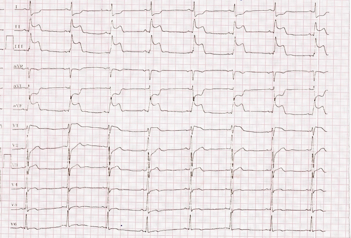
So, diagnosis of inferior wall myocardial infarction with Right ventricular infarction was made as there was ST segments elevation in lead III was more than lead II (III>II) and ST segment elevation in V1 with segment depression in V2 is highly specific of RV infarction when right sided chest leads are not available.
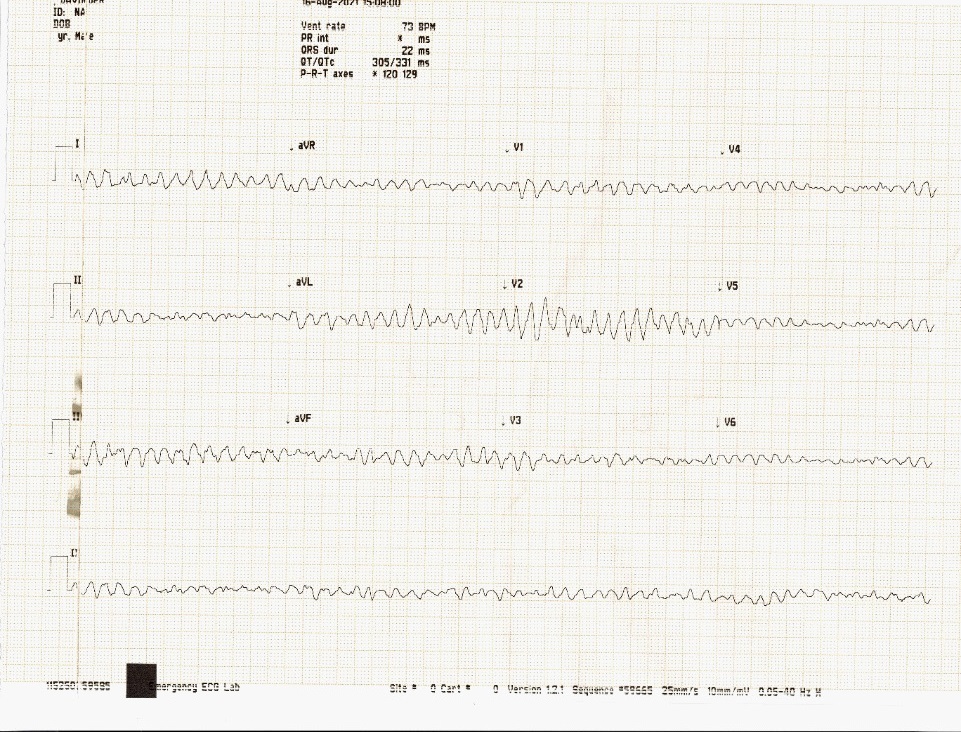
Patient developed seizure like activity while taking another ECG (Figure 2) and polymorphic broad complex ventricular arrhythmia was captured in ECG which was terminated with DC shock of 150 J. As chest pain was of more than 2 hours duration so thrombolytics were administered and Post thrombolysis ECG was repeated (Figure 3).
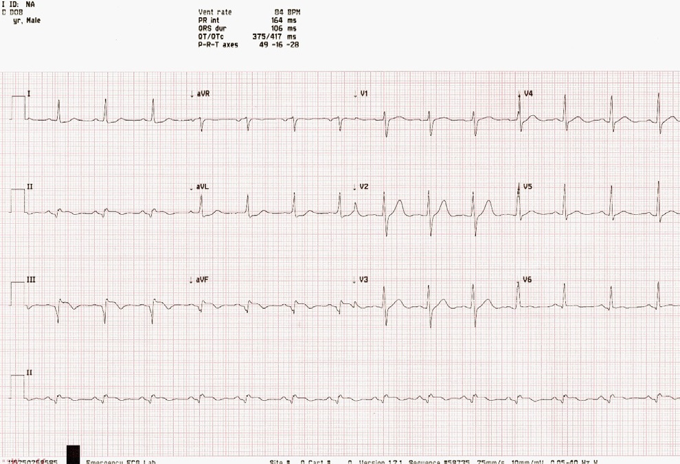
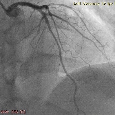
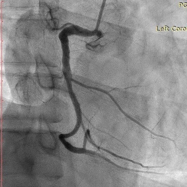
Following thrombolysis there was complete resolution of chest pain with more than 50% resolution ST segment elevation in inferior leads and in lead V1 suggestive of successful thrombolysis. Patient underwent coronary angiography which was showing normal Right coronary artery, LAD and LCX artery (Figure 4 & 5)
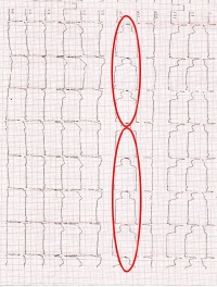
The interesting finding present here in this ECG is that ST segment elevation in aVF and ST segment depression in aVL combinedly giving appearance of a “DEAD MAN SIGN” (dead man lying on ground). This ECG sign is associated with total or near total occlusion of right coronary artery
Discussion
Dead man sign of ECG is suggestive of total or near total occlusion of proximal right coronary artery with course of bradyarrhythmia and tachyarrhythmia (supraventricular or ventricular tachyarrhythmias). In this case patient presented with bradycardia with the heart rate of 55/min but later he developed broad complex ventricular arrhythmia which is typically associated with dead man sign when seen with inferior wall myocardial infarction [4].
Patient received thrombolytics as presentation was more than 2 hours. Following thrombolysis ST segment elevation subsided more than 50%. However proximal right coronary artery obstruction was normal on coronary angiography as patient already received thrombolytics with resolution of ST segment elevation following thrombolysis is suggestive of successful thrombolysis.
Conclusions
In conclusion, this case demonstrates inferior wall myocardial infarction with RV myocardial infarction with the heart rate of 55/min later on patient developed polymorphic broad complex Ventricular tachyarrhythmia demonstrating the usual presentation seen with the dead man sign in ECG with inferior wall myocardial infarction.
References
- Shriki JE, Shinbane JS. Identifying, characterizing, and classifying congenital anomalies of the coronary arteries. Radiographics. 2012; 32: 453-68.
- Aydin F, Turgay Yildirim O. Acute Inferior Myocardial Infarction Caused by Lightning Strike. Prehosp Disaster Med. 2018; 33: 658-659.
- Konstam MA, Kiernan MS, Bernstein D, et al. Evaluation and Management of Right-Sided Heart Failure: A Scientific Statement From the American Heart Association. Circulation. 2018; 15: 2021.
- Ahamad Tramboo N. A New ECG Sign. Int J Clin Med Imaging. 20162021; 3: 3.