
Journal of Clinical Images and Medical Case Reports
ISSN 2766-7820
Case Report - Open Access, Volume 2
Acute pulmonary embolism: Tell tale signs
Mayank Yadav
PGIMER, Advanced Cardiac Center, Sector 12, Chandigarh 160012, India.
*Corresponding Author: Mayank Yadav
PGIMER, Advanced Cardiac Center, Sector 12,
Chandigarh 160012, India.
Email: mayank001.my@gmail.com
Received : Sep 27, 2021
Accepted : Nov 18, 2021
Published : Nov 25, 2021
Archived : www.jcimcr.org
Copyright : © Yadav M (2021).
Citation: Yadav M. Acute pulmonary embolism: Tell tale signs. J Clin Images Med Case Rep. 2021; 2(6): 1425
Introduction
Acute Pulmonary embolism is one of the major preventable causes of in hospital mortality. It is commonly seen in ICU setting in chronic bed ridden patients [1]. It has wide spectrum of clinical presentation ranging from asymptomatic stage to severe hemodynamic decompensation so diagnosis requires high degree of suspicion. In majority of cases detailed history and physical examination along with ECG and 2D transthoracic echocardiography is enough the diagnosis. CT pulmonary angiography is done to confirm the diagnosis or when diagnosis is not possible by other non-invasive tests. A 45-year-old army person presented to ER with breathlessness for 3 days. There was no complaint of chest pain, giddiness, palpitation, swelling or pain of legs. Patient does not have any other comorbidities.
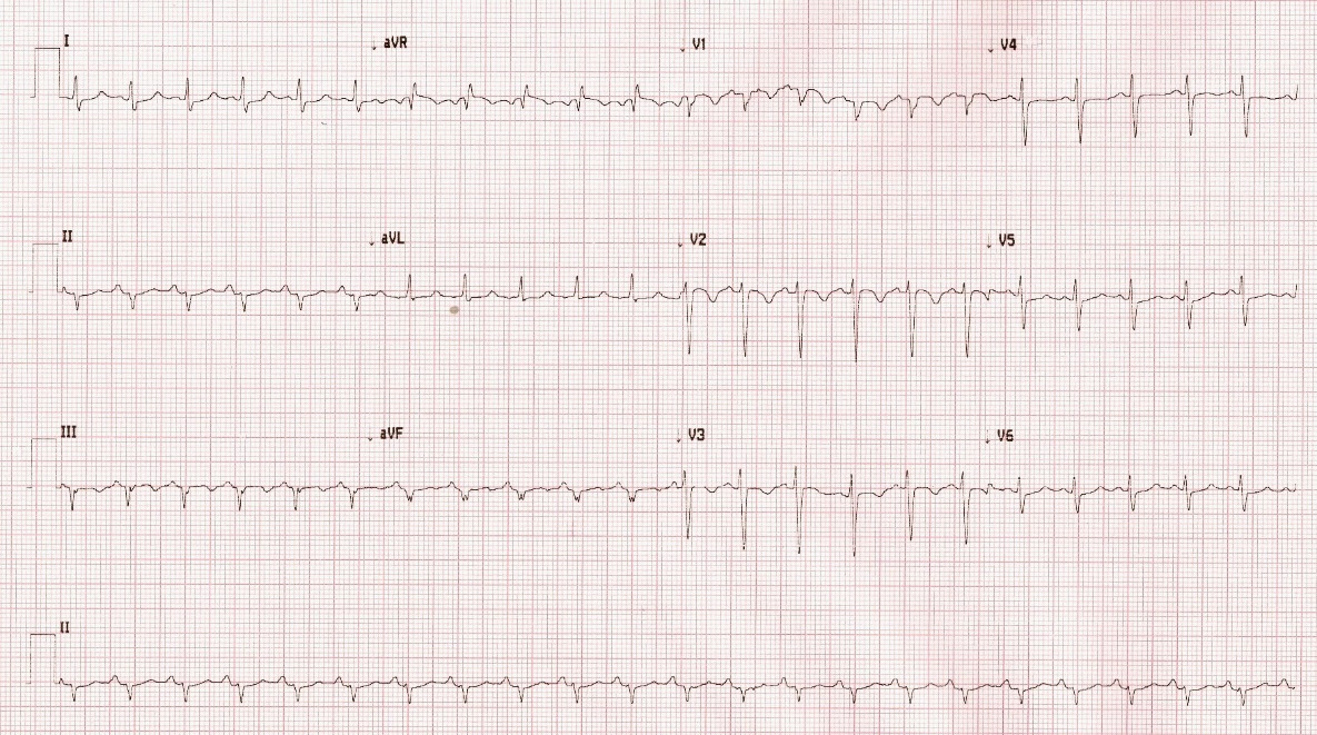
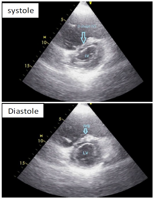
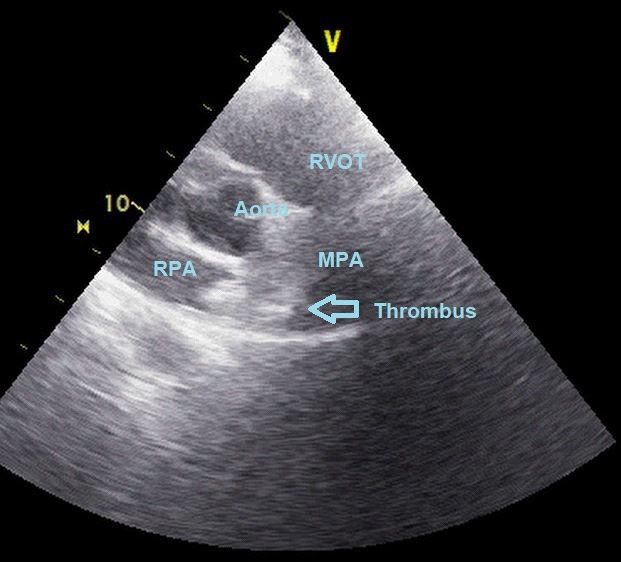
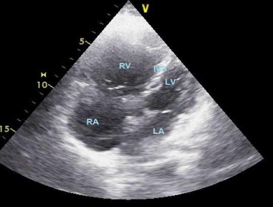
At presentation patient was having heart rate of 140/min with blood pressure of 110/84 mmHg and saturation 88% at room air and 95% with oxygen by mask. Basic investigations including ECG and echocardiography were done at presentation. 12 lead ECG (Figure 1) showing narrow complex sinus tachycardia with S1Q3T3 pattern and T wave inversion from V1-V3. 2D transthoracic echocardiography was done showing (Figure 2-4) classical features of pulmonary embolism including dilated right atrium and right ventricle, Moderate tricuspid regurgitation with elevated RVSP, dilated main pulmonary artery, Flattening of intraventricular septum during systole (D shape), Large thrombus in right pulmonary artery.
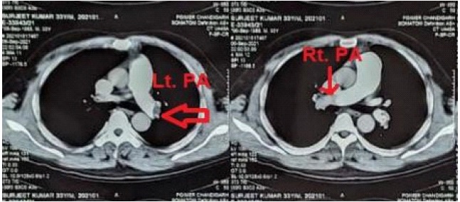
Compression USG of both lower limbs was done was negative for venous thrombosis. To conform the diagnosis of pulmonary embolism and to rule out other causes of breathlessness. CT pulmonary angiography was performed (Figure 5) which was showing the thrombus in both right and left pulmonary arteries. So, diagnosis of acute pulmonary embolism was made with no concurrent DVT.
Patient was treated with anticoagulants and with oxygen support and discharged on oral anticoagulation after one week. Diagnosis of pulmonary embolism requires high degree of suspicion as it have wide spectrum of clinical presentation. Most of the time detailed history, physical examination along with ECG and 2D echocardiography is enough to make the diagnosis of pulmonary embolism but CT pulmonary angiography is the investigation of choice as it easily available, noninvasive and high sensitivity and specificity and confirm the diagnosis [2]. Most common ECG finding in pulmonary embolism is sinus tachycardia seen in 40% of patients [1]. Other classical ECG findings include S1Q3T3, T wave in inversion in right precordial leads, incomplete or complete right bundle branch block. 2D echocardiography is noninvasive, bedside available attractive diagnostic tool. It can help in diagnosis of pulmonary embolism and can diagnose other causes of hemodynamic instability [3].
Common 2D echocardiographic signs includes acute dilatation of right atrium and right ventricle, presence of high pulmonary artery systolic pressure measured as elevated RVSP from tricuspid regurgitation jet, presence of thrombus can be visualized in main or branch pulmonary artery in addition to dilated pulmonary artery, Mc Connell sign and 60/60 sign. This case illustrates typical tell-tale signs of pulmonary embolism with history dyspnea and classical ECG and echocardiographic signs which finally confirmed with CT pulmonary angiography.
Conclusion
To conclude pulmonary embolism is common condition seen in emergency department with high mortality and morbidity requires early diagnosis and prompt treatment. Diagnosis can be made with detailed history and classical signs on ECG and 2D echocardiography. CTPA can be done when diagnosis is doubtful to confirm the diagnosis.
References
- Konstantinides SV, Meyer G, Becattini C, Bueno H, Geersing G-J, et al. 2019 ESC Guidelines for the diagnosis and management of acute pulmonary embolism developed in collaboration with the European Respiratory Society (ERS). Eur Heart J. 2020; 41: 543–603.
- Adibi A, Nouri S, Moradi M, Shahabi J. Clinical and echocardiographic findings of patients with suspected acute pulmonary thromboembolism who underwent computed tomography pulmonary angiography. J Res Med Sci Off J Isfahan Univ Med Sci. 2016; 21: 118.
- Lee J-H, Park J-H. Role of Echocardiography in Patients With Acute Pulmonary Thromboembolism. J Cardiovasc Ultrasound. 2008; 16: 9.