
Journal of Clinical Images and Medical Case Reports
ISSN 2766-7820
Case Report - Open Access, Volume 2
Unilateral colonization of fungal infection presenting as renal mass in young patient: A case report
Ayana Sarangi1; Biswaranjan Paital2*
1 Department of Zoology, College of Basic Science and Humanities, Odisha University of Agriculture and Technology, Bhubaneswar-751003, India.
2 Redox Regulation Laboratory, Department of Zoology, College of Basic Science and Humanities, Odisha University of Agriculture and Technology, Bhubaneswar-751003, India.
*Corresponding Author: Biswaranjan Paital
Redox Regulation Laboratory, Department of
Zoology, College of Basic Science and Humanities,
Odisha University of Agriculture and Technology,
Bhubaneswar-751003, India.
Email: biswaranjanpaital@gmail.com
Received : Oct 13, 2021
Accepted : Nov 26, 2021
Published : Dec 03, 2021
Archived : www.jcimcr.org
Copyright : © Paital B (2021).
Abstract
Getting a sufficient quantity of sleep is an important aspect of maintaining a healthy sleep pattern. A good night-sleep is the natural cure for all health problems. Sleeping disorders affect people of all ages, although adults (18-60) are the most common sufferers these days. If a person does not get enough sleep during the day, he or she may develop major health problems such as high blood pressure, high sugar, and drowsiness most of time. As a result, the person may develop Parkinson’s disease, Alzheimer’s disease, or insomnia, among other ailments. The sleep cycle is normally divided into two stages i.e. NON REM and REM. Depending on the age category, the entire cycle takes between 7 - 10 hours to complete. The REM stage will take 90 minutes to complete. This stage is regarded as the most powerful stage of the sleep cycle. This aids in improving one’s ability to memorise information. An experiment using a mouse as a model organism has shown that REM sleep can help a person retain things for a long time even when brain activity is silenced, whereas optogenetically manipulating the brain can help patients to forget events that occurred before sleep. To obtain optimal health, a person needs to take adequate sleep on a daily basis.
Keywords: hypothalamus; memory; neuronal modulation; optogenetic activity; sleep; synapse.
Citation: Sarangi A, Paital B. Association between REM sleep and strengthening memory: A mini review. J Clin Images Med Case Rep. 2021; 2(6): 1451.
Introduction
A good sleep is an important aspect to maintain circadian rhythm of our body. A person should undergo both REM and NON REM sleep which will take about 7-9 hrs in adult and in infant it takes about 10-11 hrs. Hypothalamus (part of brain) controls the cycle. Endocrine gland is also equally responsible to regulate circadian rhythm. Memory has a strong connection to sleep cycle. If the person is sleep deprived then it is very rare that he/ she can recollect the things from near past. As a result of sleeping for long hours (adult-more than 9 hrs and childrenmore than 11 hrs) then they may face sleep disorder. Synaptic connection between neurons helps to release neurotransmitters and regulate the action potential with the help of different ion channels, which helps us to boost our potential of memorizing the incidents. This is more active during sleep that is in REM stage.
Brain and memory
Brain assembles the information so that it has meaning for us, and can store the information in form of memory. It controls functioning of organs, movement of organs, thoughts, memory and speech. The nerve cells, non-neuronal cells (which help to maintain neurons and brain health), and small blood arteries are all found in the grey matter and white matter of the brain [1]. The brain has a number of specialised sections that collaborate. A person’s thoughts and voluntary movements are controlled by the cortex (the outermost layer), The brain stem (the part of the brain that connects the spinal cord to the rest of the brain) is involved in gas exchange and sleep. The basal ganglia (brain’s core) coordinate information between different parts of the brain. The cerebellum (the base and back of the brain) helps to balance and coordinate communications as shown in Figure 1.
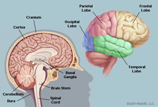
In the brain, there are several lobes. The frontal lobes are responsible for problem solving as well as motor neuron function. Sensory nerves, handwriting, and body posture are all managed by the parietal lobes. Memorize and stimulatory actions are two functions of the temporal lobes. Visualization is aided by the occipital lobes as shown in Figure 1.
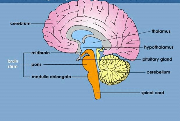
Coronal section of brain
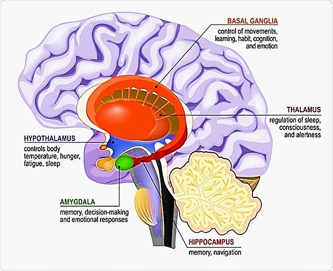
As can be seen in figure 2, the hypothalamus is in charge of sleep regulation. Melaton in is produced by the pituitary gland that aids sleep at night. The hippocampus is in charge of memory formation. Throughout one's life, the cells of a particular region divide. Dentate gyrus is a unique neuron that is present.
Synapse
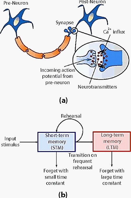
Neurons are the cells of the nervous system. They are divided into three sections as shown in Figure 3. The nucleus is found in the cell body (soma). The portion of a nerve cell (neuron) that carries nerve impulses away from the cell body is known as an axon.
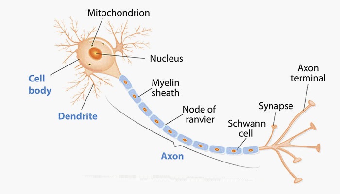
Axons are the extensions of a neuron that connect it to other neurons, muscle cells, and gland cell. Dendrites are the projections of a neuron (nerve cell) that receive and transmit signals (information) from other neurons. A synapse is a small gap at a neuron’s end that allows a signal to pass from one neuron to next.
These can be found where nerve cells meet and connect with other nerve cells. These act as keys to brain’s function especially when it comes to memory. Synapses are classified as presynaptic, postsynaptic, axodendritic, axosommatic, axoaxonal synapse, excitatory synapse, inhibitory synapse. The synapse is involved in memory formation. Synaptic cleft is linked to a neurotransmitter that strengthens the connection between two neurons. Due to a signalling process, the neurons are active at the same time. Neuronal strengthening is responsible for information storage in the form of memory. Synaptopathy is a disorder of the spinal cord, peripheral nervous system, and brain caused by synapse malfunction.
Synaptic transmission mechanism
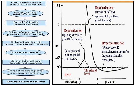
Synaptic transmission is the exchange of information across a synaptic junction, which can be accomplished through electrical, chemical, or both processes. The arrival of a nerve action potential at the axonal terminal starts a chain of events. Transmission of excitatory or inhibitory chemicals across the neuroeffector junction causes this. Synaptic vesicles are where neurotransmitter chemicals are synthesised and stored. There is synchronised release of transmitter molecules, ATP, second messenger when the nerve action potential reaches the axon terminal [6].
In the post synaptic membrane, there are two types of permeability alterations. The excitatory post synaptic potential causes a localised depolarization of the membrane by increasing the permeability of all ions mainly allows Na+ . Inhibitory post synaptic potential causes membrane hyperpolarization by increasing membrane permeability to only smaller ions such as k+ and chloride ions. Movement of ion channels is shown in Figure 4 [7,8].
Acetylcholine is a transmitter that is released at post-autonomic nerve endings in order to improve synaptic connections. Glycine, GABA (glutamine aminobutyric acid), histamine, dopamine, epinephrine, norepinephrine, and serotonin are some of the neurotransmitters involved in various physiological processes.
Memory
Memory is of two kinds. Namely long term and short term memory.
Long term memory
Long-term memory (LTM) is the stage of memory where informative knowledge is held indefinitely. It is also known as Atknison-Shiffrin memory model. Examples of long term memory– recollection of a crucial day within the distant past (early birthday, graduation, wedding, etc), and work skills you learned in your first job out of faculty. There is synaptic interaction of presynaptic and post synaptic neuron in LTP (long term potentiation). It induces by Calcium influx shown in Figure-5 and increase in glutamate sensitivity.

Short term memory
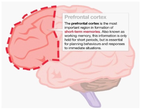
Short-term memory has been referred to as "the brain's Post-it note" because it works as a form of "scratch-pad" for temporary recollection of knowledge that is being processed at any one time. Memory is processed at prefrontal cortex as shown in Figure 6. Short-term memory Example- things like remembering where you parked your car this morning, what you had for lunch yesterday, and information from a book you just finished a few days ago. This can include anything from 18- 30 seconds to a few days' worth of activities [2].
Hippocampus
The temporal lobe, which is the primary portion of the limbic system, has a forebrain structure. Dentate gyrus, CA (cornusammonis) field CA1, CA2, CA3 Subiculum are all parts of the hippocampus. Memory, emotion, navigation, depression, and sleep are all controlled by the hippocampus, a part of the brain. Amygdala, basal ganglia, thalamus, and hypothalamus are all connected to it shown in Figure 7.
Hippocampus is a key region in the medial temporal lobe. It helps in processing the information. Through the hippocampus the short-term memory to be encoded into a long-term memory. The LTM does not remain stored in the hippocampus indefinitely. These long-term memories are crucial, and storing them in only one part of the brain is dangerous – injury to there to area would result in the loss of all of our memories. Retrograde amnesia is the loss of memories caused by damage to neurons in the hippocampus.
(a) A synapse is a connection between a pre-neuron and a post-neuron. Incoming nerve impulses from the pre-neuron cause an influx of ionic elements such as Ca2+, which causes neurotransmitters to be discharged at the synaptic connection. Short-term synaptic plasticity (STP) is the result of this, whereas numerous action potentials result in long-term potentiation (LTP). (b) Such STP and LTP mechanisms are frequently linked to the psychological model of human memory, which states that memory transitions from a short-term memory (STM) to a longterm memory (LTM) dependent on the frequency of rehearsal of the input stimulus.
Sleep and memory
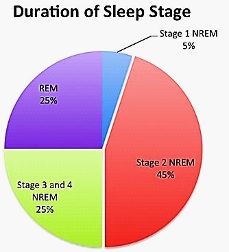
Sleep is a naturally occurring mental and physical state characterised by altered consciousness, blocked sensory activity, reduced muscle activity, reduced voluntary muscular activation during the REM stage [4], and diminished engagement with the environment. It has a stronger reaction than a comma or a state of awareness issue. It occurs over a period of time in which the body alternates between two stages of sleep: REM and NONREM. The pie chart is shown in figure-9 indicating the duration of our sleep in each stage of sleep cycle.
Lack of sleep can cause anxiety, memory loss, and a grumpy mood. A person's memory and mood tend to improve when they get enough rest. (3) The US Department of Health and Human Services set a goal to increase the percent of adults who get adequate sleep among healthy people by 2020, with the goal of improving the nation's health.
Insomnia affects about 62 % of adults worldwide. This occurs as a result of losing 1 or 2 hours of sleep a night. Because of the impact on their cognitive and motor functions, adults recognise they have been sleep deprived for one or two days.
REM sleep
Rapid eye movement is abbreviated as REM. Your eyes move around fast in a variety of directions during REM sleep, but no visual information is sent to your brain. It is characterised by muscle relaxation, eye movement, quicker breathing, and higher brain activity, among other physiological changes.
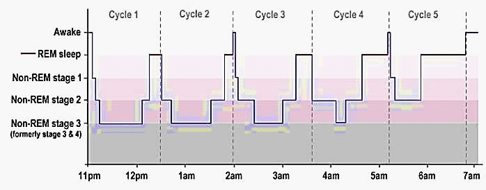
It happens roughly 90 minutes after a person falls asleep for the first time. In figure-10, the red lines indicate the time spend during REM sleep and blue line indicates NON-REM sleep. Atonia is a transient paralysis that affects the majority of people,this happens during REM stage. REM sleep is critical for brain growth, mood, memory consolidation, and dreaming. REM sleep behaviour problem is occasionally observed. Unpleasant nightmares, forceful arm and leg movement, loud noise, and so on are examples of this. This happens from time to time. Sometimes this is referred to dream enacting behaviour.
Memory during REM sleep
Dreams can be a mechanism for the brain to consolidate memory [4]. It is at this time that the brain may rearrange and analyse the event, as well as connect new and old experiences. This indicates that Body is turned off. This is something that the brain can do on its own. This is known as the activation synthesis theory. It proposes that dreams are the brain's attempt to make sense of random signals occurring during sleep. When people are taught a skill and then deprived of REM sleep, they frequently remember what they were taught.
Memory consolidation during REM sleep
Pons connects with the cerebral cortex via the thalamus. It functions as a sensory information filter and a motor control function. The information is processed by the cerebral cortex, which is a component of the brain that contributes to thinking, learning, and organising [9]. These parts of the brain that are activated during REM sleep and aid in learning and memory. Researchers discovered that infants spend 50% of their sleep time in the REM phase, indicating that they have a tremendous amount of learning potential.
Brain development during REM sleep
During infancy, the central nervous system develops. This stage assists in the development and strengthening of neuronal connections [5].
Dream
A dream may occur at any stage in one's life. The dream that we can see during REM sleep, on the other hand, is the most common and vivid. It can be nice at times and awful at others. If the dream is unpleasant, the person may be suffering from REM sleep disorder. A good sleep can lead to sharper concentration, better decision making, improved memory, ability to manage stress, better immune system, improved physical health, increased energy etc. A case study done by Rene Hen and colleagues at Columbia University found that adult mice require the stimulation of immature brain cells not just to acquire memories, but also to consolidate them during rapid eye movement sleep (Figure 11).
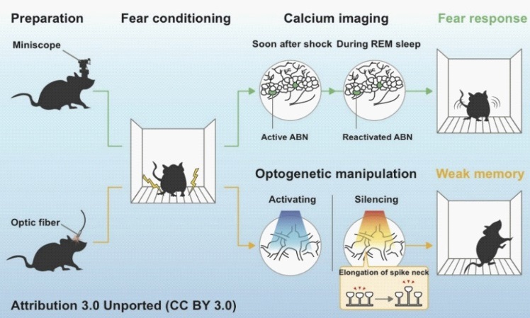
Mice were kept in a discomfort zone that is in a close box. Electrical shock is given (fear conditioning) for mice conditional learning behaviour shown in figure 11. It was noticed that deactivated neurons are silent. Mice forget all those things that they learned before sleep. This shows weak memory. This happens due to neuronal modulation.
Conclusion
In normal condition, REM sleep can help to restore the memory for a long time. Optogenetically manipulation indicates that REM sleep forgets fear conditioning memories, but restores the memories when kept undisturbed without any manipulation. Optogenetically manipulation shows that neuronal modulation can be use as a treatment for the Disease person to forget all traumatized event after a REM sleep. Therefore, a good sleep is the key behavioural adaptation to avoid many diseases associated to insomnia and associated complications.
Declarations
Acknowledgements: The figures from various internet sources with creative common attribution licence are acknowledged.
References
- Singh, Inderbir. A brief review of Techniques use in the study of Neuroanatomy”. Text book of Human Neuroanatomy (7th edition) Jaypee Brothers, New Delhi, India. 2006.
- Gold Stein,E. Bruce 1941. Cognitive psychology: connecting mind research and every day experiences 4th ed. 2015.
- Ferri R, Manconi M, Plazzi G, Bruni O, Vandi S, Montagna P, Ferini-Strambi L, Zucconi M. A quantitative statistical analysis of the submentalis muscle EMG amplitude during sleep in normal controls and patients with REM sleep behavior disorder. J Sleep Res. . 2008; 17: 89-100.
- Mutz J, Javadi AH. Exploring the neural correlates of dream phenomenology and altered states of consciousness during sleep. Neurosci Conscious. 2017: nix009.
- Dumoulin Bridi MC, Aton SJ, Seibt J, Renouard L, Coleman T, Frank MG. Rapid eye movement sleep promotes cortical plasticity in the developing brain. Sci Adv. 2015; 1: e1500105.
- Purves D, Augustine GJ, Fitzpatrick D, et al., editors. Sunderland (MA): Sinauer Associates. 2001.
- Takagi H. Roles of ion channels in EPSP integration at neuronal dendrites. Neurosci Res. 2000; 37: 167-71.
- Noga Vardi, Ling-Li Zhang. In Physiology and Pathology of Chloride Transporters and Channels in the Nervous System, 2010.
- Ahuja S, Chen RK, Kam K, Pettibone WD, Osorio RS, Varga AW. Role of normal sleep and sleep apnea in human memory processing. Nat Sci Sleep. 2018; 10: 255-269.
- Rapid eye movement sleep, WIKIPEDIA. https://en.wikipedia. org/wiki/Rapid_eye_movement_sleep
- REM Sleep. https://www.google.com/ search?q=REM+Sleep&rlz=1C1CHBD_enIN805IN805&oq=REM+ Sleep&aqs=chrome..69i57j69i59j69i59i271j69i61.159j0j4&sour ceid=chrome&ie=UTF-8
- What is REM Sleep and it’s effect. www.medicalnewstoday.com.
- Stages of REM sleep (https://www.webmd.com/sleep-disorders/sleep-101)
- Advantages of REM sleep (https://www.webmd.com/sleep-disorders/sleep-101)
- REM sleep and memory (https://www.verywellmind.com/understanding-dreams-2224258)
- Case study on mice adult born neuron(https://www.the-scientist.com/news-opinion/adult-born-neurons-strengthen-memories-while-mice-sleep-67610).
- Hall J, Hall M, Guyton and Hall Textbook of Medical Physiology. 14th Edition, eBook ISBN: 9780323640039, paperback ISBN: 9780323672801, Hardcover ISBN: 9780323597128, Imprint: Elsevier. 2020.
- Chatterjee’s CC. Human physiology (12th Edition). CBS publishers and distributors, India. 2020.