
Journal of Clinical Images and Medical Case Reports
ISSN 2766-7820
Case Report - Open Access, Volume 2
Cerebral abscesses in Marfan’s disease
Lacasse Marion1; Le Brun Cécile2*
1 Service de Médecine Interne et Maladies Infectieuses, CHRU de Tours, France.
2 Service de Bactériologie-Virologie, CHRU de Tours, France.
*Corresponding Author: Le Brun Cécile
Laboratoire de Bactériologie–Virologie CHRU de
Tours, Hôpital Bretonneau, 37000 Tours, France.
Email: c.lebrun@chu-tours.fr
Received : Oct 12, 2021
Accepted : Nov 30, 2021
Published : Dec 07, 2021
Archived : www.jcimcr.org
Copyright : © Cécile LB (2021).
Citation: Lacasse M, Le Brun C. Cerebral abscesses in Marfan’s disease. J Clin Images Med Case Rep. 2021; 2(6): 1458.
Clinical image description
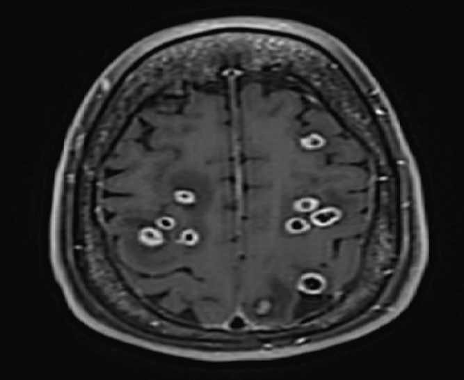
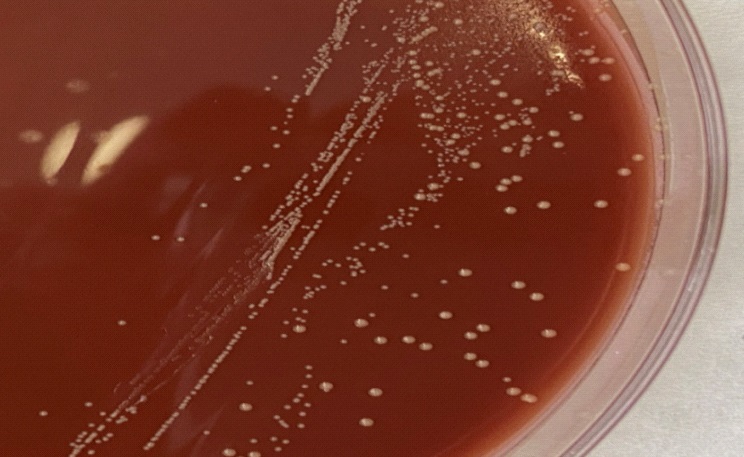
A 43-years-old woman was hospitalized with headaches progressing over the 10 preceding days and an epileptic seizure. The patient had a Marfan disease which required two cardiac surgeries - the last one in 2014 with the installation of an aortic tube and a mechanical aortic valve. A gadolinium-enhanced magnetic resonance image of the brain was performed and revealed numerous lesions, some with ring enhancement, substantial edema but without mass effect (Figure 1). Additional tests were carried out including blood cultures, transthoracic and trans-oesphageal echocardiogram, Coxiella burnetii, Bartonella spp. and Mycoplasma pneumonia serologies that remained all negative. After 48 h incubation of CT-guided brain abscess aspiration, small yellow colonies appear on chocolate blood agar and Gram staining shows small Gram-negative bacilli (Figures 2,3). Identification realized by MALDI-TOF mass spectrometry revealed Aggregatibacter aphrophilus. Blood cultures remained sterile.
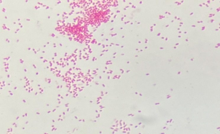
Aggregatibacter aphrophilus is a Gram negative coccobacillus (Panel C) that belongs to the HACEK group that causes 5%- 10% of infectious endocarditis in non-IV drug users. Brain abscesses are mainly described in children and most of them had cyanotic congenital heart disease as a predisposing risk factor. [1-3] .
Differential diagnosis of the brain lesions also included lymphoma, fungus, neurocysticercosis, toxoplasmosis and tuberculosis [3,4].
A PET scan has been performed and shows hypermetabolic fixation in the aortic tube. The patient therefore has endocarditis on material complicated by cerebral emboli with Aggregatibacter aphrophilus. The patient improved on cefotaxime and steroids.