
Journal of Clinical Images and Medical Case Reports
ISSN 2766-7820
Case Report - Open Access, Volume 2
PET-CT save a day in deep-seated infection
Kam Ka Ho Kevin*; Li Chun Man Timothy
Department of Medicine & Therapeutics, Prince of Wales Hospital, The Chinese University of Hong Kong, Hong Kong SAR.
*Corresponding Author: Kam Ka Ho Kevin
Department of Medicine & Therapeutics, Prince of
Wales Hospital, The Chinese University of Hong Kong,
Hong Kong SAR
Email: kkh381@ha.org.hk
Received : Oct 16, 2021
Accepted : Dec 02, 2021
Published : Dec 09, 2021
Archived : www.jcimcr.org
Copyright : © Kevin KKH (2021).
Abstract
Ventriculoperitoneal (VP) shunt infection is one of the common reasons of shunt malfunction. However, it is a well-known scenario that most patients with VP shunt infection present with non-specific complaints like fever and abdominal discomfort. Lumbar puncture analysis and blood culture could be unrevealing which may delay the diagnosis and subsequent management. Thus, the introduction of 2-[18F]-fluoro-2- deoxy-D-glucose (2-[18F]-FDG) positron emission tomography computed tomography (PET-CT) is filling the clinical gap. PET-CT is now being recognized as the imaging modality for shunt infection with moderate clinical suspicion. Our case underlined the importance of early PET-CT in pyrexia of unknown origin in identifying deep-seated infection.
Keywords: VP shunt infection; PET-CT.
Citation: Kevin K, Timothy L. PET-CT save a day in deep-seated infection. J Clin Images Med Case Rep. 2021; 2(6): 1462.
Description
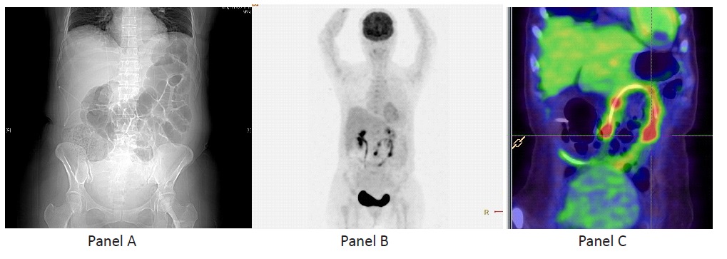
A 60-year-old female had implantation of Ventriculoperitoneal (VP) shunt a year ago after suffering from subarachnoid hemorrhage with hydrocephalus. She presented this time with colicky abdominal pain and high swinging fever. Abdominal XRay (Panel A) was suggestive of intestinal obstruction due to paralytic ileus, which was treated conservatively. However, her fever persisted despite multiple broad-spectrum antibiotics. Whole body computed tomography with contrast was unremarkable. Lumbar puncture was done in view of intermittent confusion plus ongoing fever, it showed elevated cerebrospinal fluid total protein and high white cell count but the culture was negative. Eventually 2-[18F]-Fluoro-2-Deoxy-D-Glucose (2-[18F]-FDG) Positron Emission Tomography–Computed Tomography (PET-CT) revealed a 1.1 X 2.1 X 3.1 cm hypermetabolic (SUV max 9.1) rim enhancing collection around the tip of VP shunt, confirming the diagnosis of VP shunt infection (Panel B and C). Ventriculoperitoneal shunt infections may present with vague symptoms which make the diagnosis elusive. PETCT serves an important role given its enhanced diagnostic sensitivity. The patient completely recovered after removal of the infected shunt. From multiple case studies, PET-CT has been an emerging imaging tool in diagnosing shunt related infections in case of moderate clinical suspicion when first line of diagnostic modalities could not identify sources of infection [1,2].
References
- T Rehman, M O Chohan, H Yonas. Diagnosis of ventriculoperitoneal shunt infection using [F-18]-FDG PET: A case report. J Neurosurg Sci. 2011; 55: 161-163.
- Mathew B, Purandare NC, Agrawal A, Shah S, Puranik A, et al. Progressive Hydrocephalus Due to Ventriculoperitoneal Shunt Infection: Detection With FDG PET CT. Clin Nucl Med. 2020; 45: e146-e147.