
Journal of Clinical Images and Medical Case Reports
ISSN 2766-7820
Case Report - Open Access, Volume 2
A rare association of giant congenital melanocytic nevus (bathing trunk nevus) with cryptorchidism and inguinal hernia: Case report
Mayank Kumar1; Meera luthra1*; Vimalendu Brajesh2; Rakesh Kumar Khazanchi2
1 Department of Pediatric Surgery, Medanta, The Medicity, Gurugram, Haryana, India.
2 Department of Plastic Surgery, Medanta, The Medicity, Gurugram, Haryana, India.
*Corresponding Author: Meera Luthra
Department of Pediatric Surgery, Medanta, The
Medicity, Gurugram, Haryana, India.
Email: luthra.meera@gmail.com.
Received : Oct 19, 2021
Accepted : Dec 06, 2021
Published : Dec 13, 2021
Archived : www.jcimcr.org
Copyright : © Luthra M (2021).
Abstract
Giant Congenital Melanocytic Nevus (GCMN) are rare cutaneous lesions occurring in 2,00,000 - 5,00,000 births. We are reporting a child who had left undescended testis and inguinal hernia along with GCMN. Along with orchidopexy and hernia repair, a test patch dermabrasion of nevus was done. We review our experience with this rare entity.
Citation: Kumar M, Luthra M, Brajesh V, Khazanchi R. A rare association of giant congenital melanocytic nevus (bathing trunk nevus) with cryptorchidism and inguinal hernia: Case report. J Clin Images Med Case Rep. 2021; 2(6): 1470.
Introduction
Melanocytic nevi are benign proliferations of melanocytic cells (also known as nevus cells) arranged in nests in the epidermis, dermis or in other tissue. The lesions present at birth are, by consensus, defined as congenital [1]. Giant Congenital Melanocytic Nevus (GCMN) are characterized by its extensive size and is defined as melanocytic nevus measuring more than 20 cm in its greatest dimension. They occur in approximately one in 2,00,000-5,00,000 births, more common in females [2]. The common sites of GCMN are lower back and thigh area. They are usually deeply pigmented, covered with moderate growth of hair and often there are scattered satellite lesions associated with them. They are a cause of great cosmetic concern and take a psychological toll on the patients and their family. In approximately 13.7% of cases these may undergo malignant transformation [2]. Lesions involving neck may be associated with leptomeningeal melanocytes and may produce seizures, hydrocephalus and mental changes [2]. Their association with neurocutaneousmelanosis, diffuse lipomatosis, von-Recklinghausen’s disease, vitiligo, hypertrophy of skull bones, spina bifida, meningocele and club foot have been reported [3].
Case report
We present here a case of 5-month male child, with uneventful antenatal history, presenting with a hyper pigmented skin lesion. There was no positive family history or history of consanguineous marriage.
General physical examination and systemic examination did not reveal any other abnormality.
Cutaneous examination revealed an extensive brown-black patch covering most part of the trunk, buttocks, thighs, genitals and extending upto the knee level, encircling the trunk circumferentially. A 1.5 X 1.5 cm sized patch was also seen over the right cheek. Multiple small hyper pigmented patches of varying sizes were also seen over the trunk, limbs and scalp. The skin lesions had been present since birth and had increased in size proportional to his growth. It was raised above the skin surface and was rugose, leathery with coarse hairs. Mucosal surfaces, scalphairs, nails, palms and soles were normal (Figure 1a,b,c).
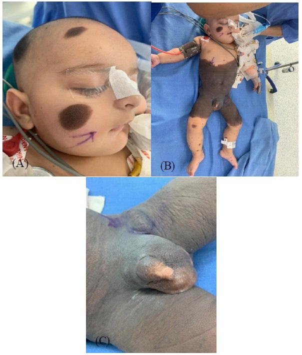
Local examination of genitals and groin revealed normal penis, palpable right testis and empty left hemiscrotum. Globular swelling with expansile cough impulse was present on left side. A small 1 X 1 cm sized mass was palpable in the left mid inguinal region. An ultrasonography was done which showed left testis in superficial inguinal pouch.
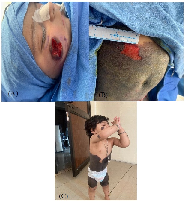
Plastic surgery consultation was sought for the pigmented lesion. The baby was operated on 27/11/19 for inguinal hernia and undescended testis. The left testis was intraoperatively found to be present in the left inguinal canal. A left herniotomy and left orchidopexy was performed first. The plastic surgery team did first stage of serial excision and closure for the lesion present on the right cheek and dermabrasion of a small patch of hyper pigmented area over the abdomen (Figure 2a,b). This was done as a test patch to see the outcome. Excised tissue was sent for histopathological examination. Post-operative period was uneventful and patient was discharged on day two of surgery.
The biopsy results were suggestive of benign nevus with congenital pattern features. On 1 year follow up, the child is growing well. The pigmented lesion responded well to the dermabrasion and the lesion on the face didn’t grow further (Figure 2c).
Discussion
Bathing suit nevi is an uncommon anomaly with a classical appearance and a spot diagnosis. Association of their congenital abnormalities has been reported. Various treatment modalities are present for GCMN like surgical excision with grafting, skin curettage, dermabrasion, tangential excision, chemical peels, Q switched and carbon-di-oxide lasers [1]. The treatment approach should be taken after considering age of patient, size and site of lesion, risk of melanoma, changes suggestive of malignancy, functional impairments and psychological impact associated with it.
50% of melanomas found in patients with GCMN occur elsewhere. Therefore, the removal of the nevus does not guarantee protection against malignancy [1,4]. The size of the lesion may be too large to allow complete excision. Tissue expanders, serial excision, skinflaps, grafts or a combination of more than one type of surgical procedure have been used.
Dermabrasion and skin curettage as treatment options are most efficient if performed in the first two to six weeks of life, when it is easier to find a cleavage plane between the upper dermis, rich in nevic cells, and the deeper dermis, relatively poor in these cells. This can be performed in one session. Tangential excision of the upper dermis may cause mild scarring, sometimes with dotted pigmentation, in infants. Chemical peels can improve the appearance of lighter GCMN or those whose nevus cells are more superficially located [1].
In our patient at 1 year follow up, the dermabrasion site was seen to have reduced pigmentation and there was no recurrence indicating the usefulness of procedure even if done at 5 months of age. Results show that dermabrasion can be a good alternative to the surgical excision as far as the cosmetic issues are concerned. It is simple procedure and has lesser side effects than surgical excision. However, the protection it provides against development of melanoma is needed to be studied. Even after complete removal of lesion regular follow up is mandatory
Ibrahim A. et al. have reported a case of a child who had ambiguous genitalia and undescended test is along with GCMN [5]. Occurrence of undescended test is and inguinal hernia with GCMN is rare and this is the only other case reported in literature to the best of our knowledge.
References
- Viana ACL, Gornitizo B, Bittencourt FV. Giant congenital melanocytic nevus. An Bras Dermatol. 2014; 89: 190.
- Keen AA, Hassan I. Giant congenital melanocytic nevus (bathing trunk nevus) in a neonate. Indian journal of Pediatr Dermatol. 2014; 15: 53-54.
- Bhagwat PV, Topkhane RS, Shashikumar BM, Noronha TM, Naidu V, et al. Giant congenital melanocytic nevus (bathing trunk nevus) associated with lipoma and neurofibroma: Report of two cases. Indian J Dermatol Venerol Leprol. 2009; 75: 495-498.
- De David M, Orlow SJ, Provost N, Marghoob AA, Rao BK, Huang CL et al. A study of large congenital melanocytic nevi and associated malignant melanomas: review of cases in the New York University Registry and the world literature. J Am Acad Dermatol. 1997; 36: 409–416.
- Ibrahim A, Ramatu A. Congenital giant melanocytic nevus with ambiguous genitalia in an 8 year old child: A rare combination. Saudi J Health Sci. 2013; 2: 214-216.