
Journal of Clinical Images and Medical Case Reports
ISSN 2766-7820
Case Report - Open Access, Volume 2
Unilateral livedo reticularis in ruptured abdominal aorta aneurysm
Daniella Rahim1*; Carsten Sauer Mikkelsen2; Peter Bjerring3; Lorenz Oppel4
1 Cand Med Resident, Department of Neurology, Aalborg University hospital, Denmark.
2 Specialist in Dermato-Venereologi, Private Practise in Dermato-venereologi, Brønderslev, Senior Research Fellow, Department of Dermato-venereology, Aalborg University hospital, Denmark
3 Specialist in Dermato-Venereologi, Clinical Professor, Dr.Med., Department of Dermato-Venereology, Aalborg University Hospital, Denmark.
4 Specialist in Neurology, Senior Hospital Physician, Clinical Proffesor, Dr.Med., Department of Neurology, Aalborg University hospital, Denmark.
*Corresponding Author: Daniella Rahim
Cand Med Resident, Department of Neurology, Aalborg University hospital, Denmark.
Email: daniellarahim14@gmail.com
Received : Oct 20, 2021
Accepted : Dec 06, 2021
Published : Dec 13, 2021
Archived : www.jcimcr.org
Copyright : © Rahim D (2021).
Abstract
Livedo Reticularis (LR) is a well-known, relatively common physical finding consisting of macular, violaceous, connecting rings that form a netlike-pattern.
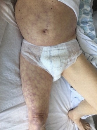
Generalized livedo reticularis is a common physiological response to cold, but may be a sign of different systemic diseases. In the present case, a 79-year male presented with unilateral live do reticularis in the right leg, secondary to ruptured abdominal aorta aneurysm.
Citation: Rahim D, Mikkelsen C, Bjerring P, Oppel L. Unilateral livedo reticularis in ruptured abdominal aorta aneurysm. J Clin Images Med Case Rep. 2021; 2(6): 1472.
Introduction
Livedo Reticularis (LR) is a net-like discoloration of the skin, due to disturbance in blood flow pattern in the superficial vessels of the skin. The low blood flow will result in higher relative amounts of deoxygenated hemoglobin, leading to a more violaceous/blueish color of the superficial capillaries [1,2]. LR is classified as either primary idiopathic or secondary to other underlying pathological conditions. Any physiological process, that impairs the blood flow can cause LR, such as cold, hyper-viscosity or systemic inflammation. Unilateral LR may also be present after local heat injury, also referred to as Erythemaabigne [2].
Case report
A 79-year male with a medical history of hypertension and prostate cancer was treated with watch-and-wait strategy. He was admitted dyspnea and fever and treated with penicillin for pneumonia. He responded with improved respiration and decreasing CRP. However, at the fifth day of admission he developed paresis of the left leg. The left leg exhibited pale skin and paresis and LR was found on the trunk and right leg (Figure 1). Furthermore, palpatory pain was registered in the lower left abdominal fossa. A normal ABCDE-status was found and blood test showed an insignificant low hemoglobin (7.2 mmol/l).
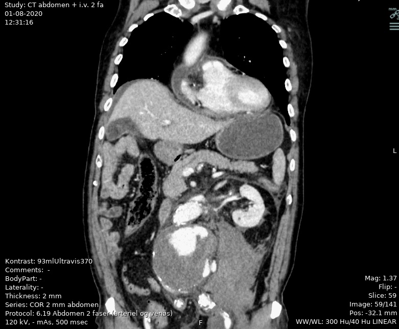
Acute CT-scan of the abdomen revealed an eight cm left-sided, ruptured infra-renal abdominal aorta aneurysm with retroperitoneal hematoma (Figure 2). The patient underwent acute abdominal surgery with insertion of a stent. Post-surgically, the patient had regained normal power in left leg and the skin colour normalized.
Discussion
LR is caused by a disturbance in the blood flow in the superficial vessels in the skin. The reduced and re-directed blood flow increases the relative amount of deoxygenized hemoglobin to oxygenized hemoglobin resulting in a prominent blue/ violet color of the visible, superficial vessels. Universal LR is a normal physiological response to cold but may also be a sign of systemic disease such as sepsis [1] and even COVID-19 [3]. Focal LR is rare, but have been presented in case reports with bilateral LR in the legs, caused by cholesterol embolisms originating from abdominal aorta aneurysms, which further caused secondary kidney failure [4].
It is speculated that the present LR was caused by activation of the sympathetic nervous system, as a response to blood loss due to the erupted aorta aneurysm. The blood flow was further obstructed to the left leg due to the retroperitoneal hematoma thereby causing paresis and pale colour of the left leg.
References
- Sajjan V, Swamy M, Lunge S, Pandit A. Livedo reticularis: A review of the literature. Indian Dermatol Online J. 2015; 6: 315.
- Gawkrodger DJ (David J., Ardern-Jones MR. Dermatology : an illustrated colour text [Internet]. 7th ed. 2020; 164.
- Manalo IF, Smith MK, Cheeley J, Jacobs R. A dermatologic manifestation of COVID-19: Transient livedo reticularis. J Am Acad Dermatol. 2020; 83: 700.
- Singh NP, Gupta AK, Kaur G. Atheroembolic renal disease. Vol. 30, Indian Journal of Nephrology. Wolters Kluwer Medknow Publications. 2020; p. 1–2.