
Journal of Clinical Images and Medical Case Reports
ISSN 2766-7820
Case Report - Open Access, Volume 2
Hyperpigmented skin lesions in a patient with severe combined immunodeficiency and adenosine deaminase gene defect
Mahnaz Jamee; Samin Sharafian*; Seyedeh Atefeh Hashemimoghaddam; Zahra Chavoshzadeh
Immunology and Allergy Department, Mofid Children’s Hospital, Shahid Beheshti University of Medical Sciences, Tehran, Iran.
*Corresponding Author: Samin Sharafian
Immunology and Allergy Department, Mofid Children’s
Hospital, Shahid Beheshti University of Medical
Sciences, Tehran, Iran.
Email: Dr.saminsharafian@sbmu.ac.ir
Received : Oct 25, 2021
Accepted : Dec 09, 2021
Published : Dec 16, 2021
Archived : www.jcimcr.org
Copyright : © Sharafian S (2021).
Keywords: skin disorder; adenosine deaminase deficiency; ADA; SCID; inborn errors of immunity.
Citation: Jamee M, Sharafian S, Hashemimoghaddam S, Chavoshzadeh Z. Hyperpigmented skin lesions in a patient with severe combined immunodeficiency and adenosine deaminase gene defect. J Clin Images Med Case Rep. 2021; 2(6): 1485.
Report
The patient was a 38-day-old male born to closely related consanguineous parents. The family history was unremarkable except for Down syndrome in his aunt.
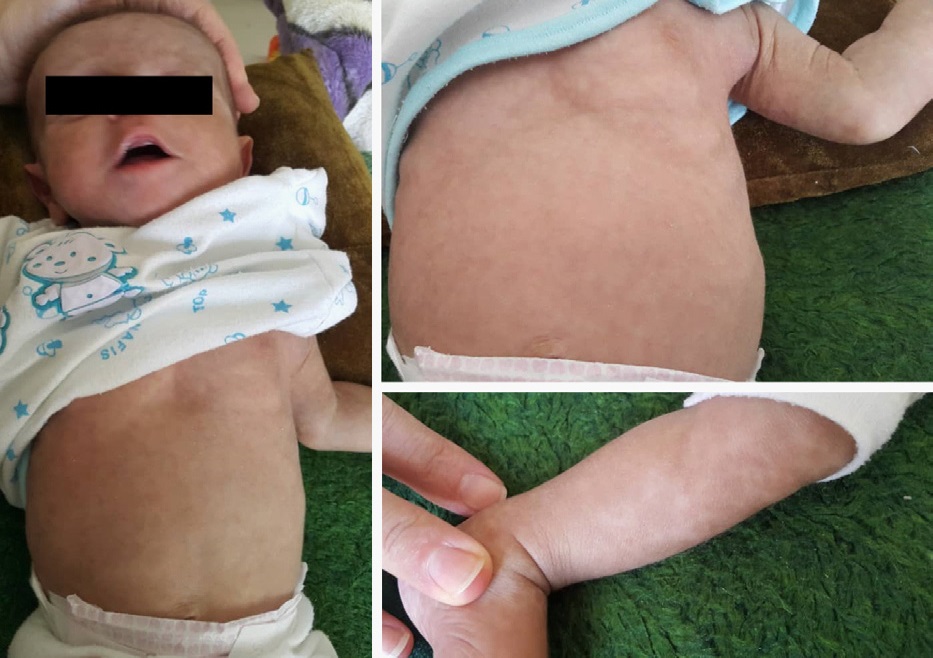
He was hospitalized due to productive cough (since about two weeks before), failure to thrive, delay in umbilical cord separation, and facial syndromic feature (low-set ear). In physical examination, generalized skin rashes were found (Figure 1). The hyperpigmented patchy rash involved the face, trunk, and extremities, with no scaling or itching, and had urticaria-like properties. The skin biopsy reported nonspecific dermal fibrosis.
In chest X-ray thymus shadow was absent. The abdominal ultrasound was normal. In laboratory evaluation, leukopenia (1500 cell/uL), low CD3+ T cells (279 cell/μL), low CD4+ T cells (139 cell/μL), low CD8+ T cells (126 cell/μL), reverse CD4+/ CD8+ ratio (1.1), low CD19+ T cells (129 cell/μL), low CD27+ memory B cells on CD19+ gate (22.22 cell/μL) and lymphocyte gate (3.22 cell/μL), and low CD16 (33.24 cell/μL) and CD56 (3.31 cell/μL) NK cells, and hypogammaglobulinemia were identified. The bone marrow aspiration had no evidence of malignancy or maturation arrest. The Nitroblue Tetrazolium (NBT) test was normal. The Lymphocyte Transformation Test (LTT) in response to mitogens and pathogens was abnormal (PHA: 1.0, BCG: 1.0). The TREC and KREC copy numbers were 134 and 945 per 106 cells, below the normal range for age.
He was diagnosed with T- B+ NK+ Severe Combined Immunodeficiency (SCID) and discharged with Immunoglobulin Replacement Therapy (IGRT) and G-CSF. The genetic study showed a homozygous missense pathogenic variant in exon 7 of the ADA gene (c.646G>A, p.Gly216Arg).
Three months later, while he was a candidate for bone marrow transplantation, he developed an axillary abscess and axillary BCG lymphadenitis. He was referred to our center for abscess drainage and was started on anti-tuberculosis treatment, however, his clinical condition deteriorated and he died following severe Pneumocystis jirovecii pneumonia, respiratory failure, and eventually septic shock.
The Adenosine Deaminase (ADA) deficiency is a monogenic inborn error of immunity characterized by SCID phenotype and mutations in the ADA gene. Along with previous reports of atopic dermatitis and dermatofibrosarcoma protuberans in ADA-SCID patients [1-3], we aim to highlight the cutaneous manifestation of ADA-SCID patients, attention to which can provide early diagnosis and a more favorable outcome.
Consent for publication: Written informed consent was obtained from parents for the participation and publication.
References
- Notarangelo LD, Stoppoloni G, Toraldo R, Mazzolari E, Coletta A, Airò P, et al. Insulin-dependent diabetes mellitus and severe atopic dermatitis in a child with adenosine deaminase deficiency. European journal of pediatrics. 1992; 151: 811-814.
- Cowen EW, Pichard DC, Garabedian E, Miettinen M. MedallionLike Dermal Dendrocytic Hamartoma, Dermatofibrosarcoma Protuberans, and Adenosine Deaminase-Deficient Severe Combined Immunodeficiency. Pediatric dermatology. 2016; 33: 359- 360.
- Wahjudi TD, Kutzner H, Bleeke M, Hoeger PH. Multicentric dermatofibrosarcoma protuberans in a child with severe combined immunodeficiency due to adenosine deaminase deficiency. Pediatric dermatology. 2021; 38: 875-878.