
Journal of Clinical Images and Medical Case Reports
ISSN 2766-7820
Case Report - Open Access, Volume 2
Pulmonary papillary adenoma: A rare benign tumor of the lung
Seongsik Bang; Seungyun Jee; Hwangkyu Son; Hyunsung Kim; Seung Sam Paik*
Department of Pathology, Hanyang University Seoul Hospital, Hanyang University College of Medicine, Seoul, Korea.
*Corresponding Author: Seung Sam Paik
Department of Pathology, Hanyang University College
of Medicine, 222, Wangsimni-ro, Seongdong-gu, Seoul
04763, South Korea.
Email: sspaik@hanyang.ac.kr
Received : Oct 25, 2021
Accepted : Dec 09, 2021
Published : Dec 16, 2021
Archived : www.jcimcr.org
Copyright : © Paik SS (2021).
Abstract
A lung mass found incidentally in a young woman was diagnosed with Pulmonary Papillary Adenoma (PPA). It is important to understand the characteristic pathologic features of PPA to differentiate it from other lung tumors.
Citation: Bang S, Jee S, Son H, Kim H, Paik S, et al. Pulmonary papillary adenoma: A rare benign tumor of the lung. J Clin Images Med Case Rep. 2021; 2(6): 1487.
Report
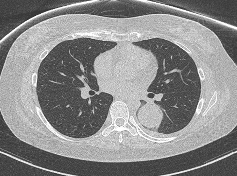
A 25-year-old woman visited the hospital to evaluate the lung mass incidentally found on a chest X-ray during the health examination at the local clinic. The computed tomography demonstrated a roundmass in the superior segment of the left lower lobe (Figure 1). Since there was no evidence of distant metastasis, she underwent lobectomy. Intraoperative consultation was performed, and we answered that non-small cell carcinoma should be considered.
On microscopic examination, the tumor was predominantly composed of tubulopapillary structures with loose myxoid fibrovascular cores lined by a single layer of cuboidal epithelium (Figure 2). The lining tumor cells lacked nuclear atypia or mitoses. Therefore, we finally diagnosed this tumor with Pulmonary Papillary Adenoma (PPA). After the final diagnosis was made, touch preparation slides were reviewed. The smears showed tight clusters with high cellularity in a myxoid background. Most cellular clusters showed cohesive sheets of uniform cells (Figure 3). After the operation, the patient is doing well without any evidence of recurrence or metastasis for two years.
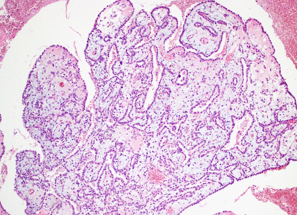
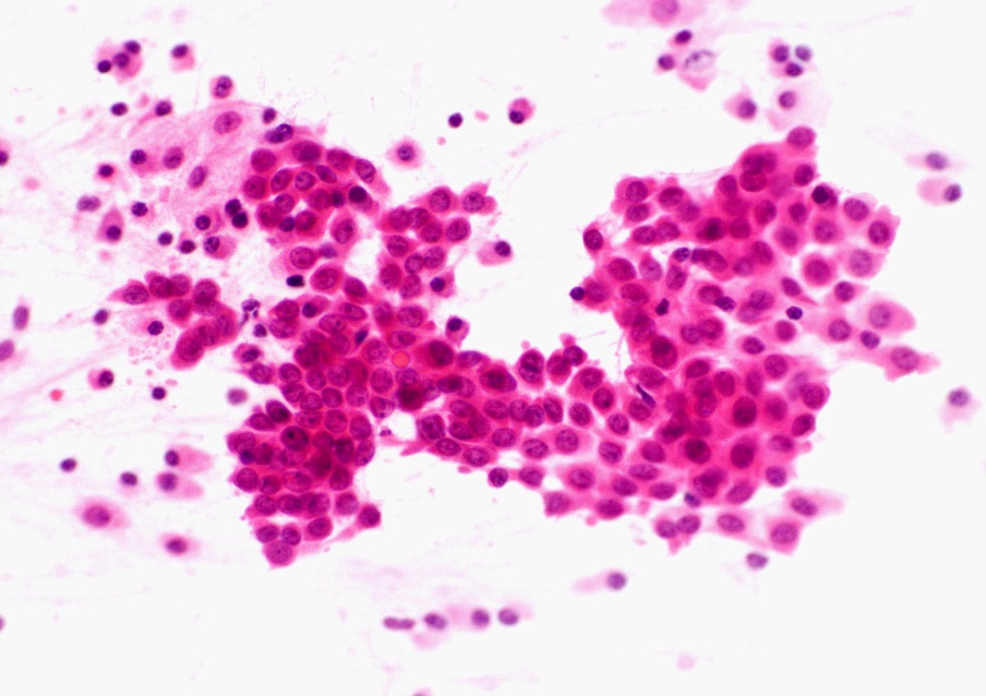
PPA is a distinct benign tumor of the lung and has a very rare incidence [1]. The histogenesis of PPA is believed to be derived from the primitive multipotent respiratory epithelium [2]. PPA shows the prominent papillary structures with fibrovascular stroma. The lining tumor cells are usually a single layer of cuboidal epithelial cells showing minimal cytological atypia [3]. On cytological examination, PPA shows high cellularity and cohesive sheets of uniform tumor cells reminiscent of type II pneumocytes [3,4]. The tumor cells show a moderate amount of eosinophilic cytoplasm. The nuclei demonstrate fine chromatin and minimal atypia with the inconspicuous nucleolus. Although the incidence is very rare, PPA should be considered as a differential diagnosis of lung tumors. Therefore, understanding the pathologic features probably helps confirm the diagnosis.
References
- Zhou P, Yu W, Wang L, Xia Q, Chen K. Retrospective study of clinical and pathologic features of pulmonary papillary adenoma. A rare tumor and 15 cases report. Medicine. 2020; 99: 44.
- Lin X-Y, Han Q, Wang E-H, Zhang Y. Pulmonary papillary adenoma presenting in central portion: A case report. Diagnostic Pathology. 2015; 10: 190.
- Frey A, Alatassi H, Wiese TA, Fraig M, Yang X. Cytomorphologic findings and differential diagnosis of pulmonary papillary adenoma. A case report and literature review. Diagn Cytopathol. 2016; 44: 543-547.
- Minami Y, Morishita Y, Yamamoto T, Iijima T, Fukasawa M, Ishikawa S, et al. Cytologic characteristics of pulmonary papillary adenoma. A case report. Acta Cytol. 2004; 48: 243-248.