
Journal of Clinical Images and Medical Case Reports
ISSN 2766-7820
Case Report - Open Access, Volume 2
Connatal cyst in a 20 year old girl
Wilson Bizimana*; Wais A Amarkak; Meryem Fikri; Mohammed Jiddane; Firdaous Touarsa
Department of Neuroradiology, Hospital of Specialities, Rabat, Morocco.
*Corresponding Author: Wilson Bizimana
Department of Neuroradiology, Hospital of
Specialities, Rabat, Morocco.
Email: wilson.bizimana@gmail.com
Received : Nov 03, 2021
Accepted : Dec 20, 2021
Published : Dec 27, 2021
Archived : www.jcimcr.org
Copyright : © Biziman W (2021).
Citation: Bizimana W, Amarkak WA, Fikri M, Jiddane M, Touarsa F. Connatal cyst in a 20 year old girl. J Clin Images Med Case Rep. 2021; 2(6): 1514.
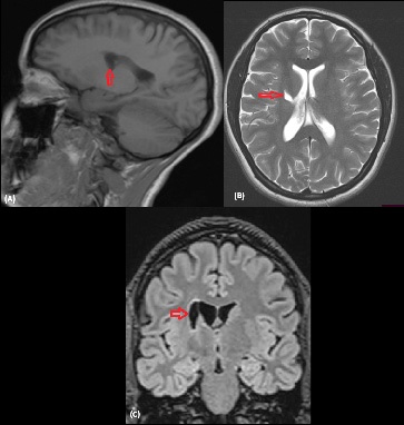
Description
Connatal cyst also known as frontal horn cysts, are cystic areas adjacent to the superolateral margins of the body and frontal horns of the lateral ventricles and are believed to represent a normal variant [1]. The other name is coarctation of the lateral ventricles or frontal horn cysts. Sometimes unilateral or bilateral, the incidence of this condition is between 0, 4 and 0, 9% [2]. Periventricular cysts are a common finding in neonatal cranial imaging. They are noted usually in antenatal period and typically disappear over the first year of the life [3]. The etiology has been debated varying from being a normal variant, to secondary to a viral infection, chromosomal anomaly, post-ischemic or hemorrhagic event [1,4]. They have been shown to regress on follow-up imaging when demonstrates in children under 2 months of age [3]. Our case demonstrates an exceptional situation of a persistent of the cyst in a 20-year-old -girl.
References
- S Unger, S Salem, L Wylie and V Shah, Newborn frontal horn cysts: Cause for concern? Journal of Perinatology. 2011; 31: 98- 103.
- Vijeyaletchimi N, Tan YR, Lee SL, Tan WC, Tan LK, Tan HK Connatal cyst: Antenatal findings and differential diagnoses. Apeksha Chaturvedi. 2017.
- C L Scelsi, T A Rahim, J A Morris, G J Kramer, B C Gilbert, and S E For seen. The Lateral Ventricles: A Detailed Review of Anatomy, Development, and Anatomic Variations. Am J Neuroradiol. 2020.
- ZY J Tan, P Naidoo, N Kenning, Ultrasound and MRI features of conatal cysts: Clinico radiological differentiation from other supratentorial periventricular cystic lesions. The British Journal of Radiology. 2010; 83: 180-183.