
Journal of Clinical Images and Medical Case Reports
ISSN 2766-7820
Case Report - Open Access, Volume 2
Right circumflex aorta: An extremely rare aortic arch anomalies
Towashiraporn K1*; Wasinrat J2
1 Her Majesty Cardiac Center, Faculty of Medicine Siriraj Hospital, Mahidol University, Bangkok, Thailand.
2 Department of Radiology, Siriraj Hospital, Mahidol University, Bangkok, Thailand.
*Corresponding Author: Towashiraporn K
Her Majesty Cardiac Center, Faculty of Medicine Siriraj
Hospital, Mahidol University, Bangkok, Thailand.
Email: korakoth.tow@mahidol.ac.th
Received : Nov 16, 2021
Accepted : Jan 06, 2022
Published : Jan 13, 2022
Archived : www.jcimcr.org
Copyright : © Towashiraporn K (2022).
Keywords: coronary angiography; aortography; computed tomography angiography; congenital anomalies of the aortic arch.
Abbreviations: CAG: Coronary Angiography; CTA: Computed Tomography Angiography.
Citation: Towashiraporn K, Wasinrat J. Right circumflex aorta: An extremely rare aortic arch anomalies. J Clin Images Med Case Rep. 2022; 3(1): 1558.
Clinical image description
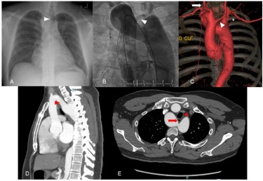
A 38-year-old Asian man was referred to our hospital due to a history of decompensated heart failure. Transthoracic echocardiography demonstrated a left ventricular ejection fraction of 29%. The chest radiograph demonstrated an increased cardiothoracic ratio. There was right paratracheal soft tissue density with compression on the right side of the trachea (Figure A, Arrowhead).
We performed coronary angiography (CAG) via the right femoral approach that revealed non-significant coronary artery disease. Regarding the difficulty of performing CAG, we did the aortography that demonstrated anomalous aortic arch. We also identify the left common carotid artery arising from the ascending aorta (Figure B, Arrowhead). 3D reconstruction of computed tomography angiography revealed left common carotid artery was the first branch that arose from the ascending aorta (Figure C, Arrowhead), followed by the right carotid artery (Figure C, Arrow), the right subclavian, and the aberrant left subclavian arteries (Figure C, Asterisk), respectively. The right arch passed between the superior vena cava and the right side of the trachea and esophagus, crossing over the right main bronchus to join the descending aorta (Figure D, Arrowhead). Mid-esophagus (Figure E, Arrow) and trachea (Figure E, Arrowhead) had anterior displacement by the pressured effect of the aberrant left subclavian artery. Suggestive of right aortic arch with circumflex aorta and aberrant left subclavian artery.
Circumflex right aortic arch is an extremely rare anomaly [1]. Surgical correction is the treatment of choice in symptomatic tracheal or esophageal compression [2,3].
References
- Pandey NN, Sharma A, Shaw M, Kumar S. Circumflex retroesophageal right aortic arch: Rare differential of mediastinal widening. BMJ Case Rep. 2018.
- Bleakney CA, Zafar F, Fraser CD. Circumflex right aortic arch with associated hypoplasia and coarctation: Repair by aortic arch advancement and end-to-Side anastomosis. Ann Thorac Surg. 2011; 91: 624–626.
- Sett SS, Lafaro RJ, Babu SC. Repair of circumflex aortic arch in an adult. Ann Thorac Surg. 2017; 104: e139–141.